2QKV image
Deposition Date 2007-07-11
Release Date 2007-11-06
Last Version Date 2024-02-21
Entry Detail
PDB ID:
2QKV
Title:
Crystal Structure of the C645S Mutant of the 5th PDZ Domain of InaD
Biological Source:
Source Organism:
Method Details:
Experimental Method:
Resolution:
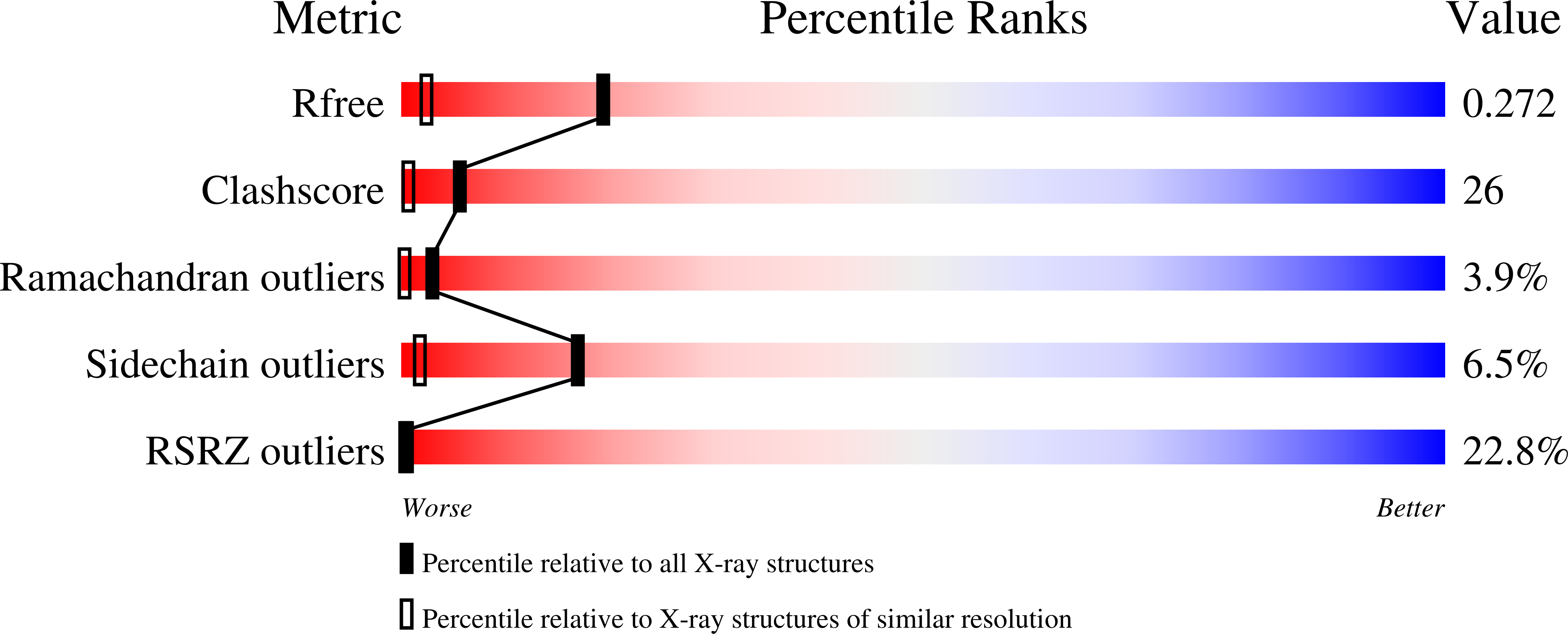
1.55 Å
R-Value Free:
0.26
R-Value Work:
0.24
R-Value Observed:
0.24
Space Group:
P 43 3 2
Macromolecular Entities
Polymer Type:polypeptide(L)
Molecule:Inactivation-no-after-potential D protein
Gene (Uniprot):inaD
Mutations:C645S
Chain IDs:A, B
Chain Length:96
Number of Molecules:2
Biological Source:Drosophila melanogaster
Primary Citation
Dynamic Scaffolding in a G Protein-Coupled Signaling System.
Cell(Cambridge,Mass.) 131 80 92 (2007)
PMID: 17923089 DOI: 10.1016/j.cell.2007.07.037

Abstact

The INAD scaffold organizes a multiprotein complex that is essential for proper visual signaling in Drosophila photoreceptor cells. Here we show that one of the INAD PDZ domains (PDZ5) exists in a redox-dependent equilibrium between two conformations--a reduced form that is similar to the structure of other PDZ domains, and an oxidized form in which the ligand-binding site is distorted through formation of a strong intramolecular disulfide bond. We demonstrate transient light-dependent formation of this disulfide bond in vivo and find that transgenic flies expressing a mutant INAD in which PDZ5 is locked in the reduced state display severe defects in termination of visual responses and visually mediated reflex behavior. These studies demonstrate a conformational switch mechanism for PDZ domain function and suggest that INAD behaves more like a dynamic machine rather than a passive scaffold, regulating signal transduction at the millisecond timescale through cycles of conformational change.

Legend

Protein

Chemical

Disease

Primary Citation of related structures