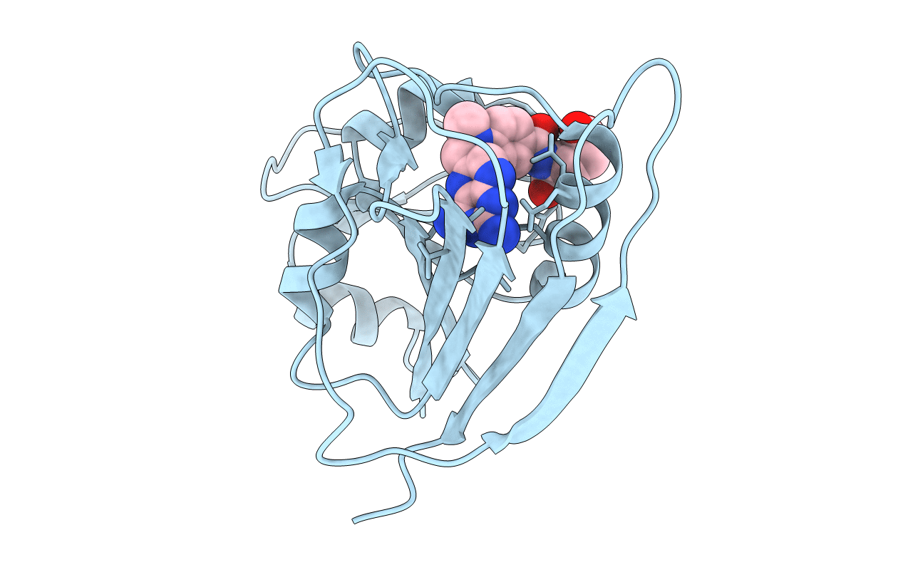
2QK8 image
Deposition Date 2007-07-10
Release Date 2007-08-28
Last Version Date 2023-08-30
Entry Detail
PDB ID:
2QK8
Keywords:
Title:
Crystal structure of the anthrax drug target, Bacillus anthracis dihydrofolate reductase
Biological Source:
Source Organism:
Method Details:
Experimental Method:
Resolution:
2.40 Å
R-Value Free:
0.27
R-Value Work:
0.24
R-Value Observed:
0.24
Space Group:
C 1 2 1
Macromolecular Entities
Polymer Type:polypeptide(L)
Molecule:Dihydrofolate reductase
Gene (Uniprot):dfrA
Chain IDs:A
Chain Length:162
Number of Molecules:1
Biological Source:Bacillus anthracis str.
Ligand Molecules
Primary Citation
Crystal structure of the anthrax drug target, Bacillus anthracis dihydrofolate reductase.
J.Med.Chem. 50 4374 4381 (2007)
PMID: 17696333 DOI: 10.1021/jm070319v

Abstact

Spores of Bacillus anthracis are the infectious agent of anthrax. Current antibiotic treatments are limited due to resistance and patient age restrictions; thus, additional targets for therapeutic intervention are needed. One possible candidate is dihydrofolate reductase (DHFR), a biosynthetic enzyme necessary for anthrax pathogenicity. We determined the crystal structure of DHFR from B. anthracis (baDHFR) in complex with methotrexate (MTX; 1) at 2.4 Angstrom resolution. The structure reveals the crucial interactions required for MTX binding and a putative molecular basis for how baDHFR has natural resistance to trimethoprim (TMP; 2). The structure also allows insights for designing selective baDHFR inhibitors that will have weak affinities for the human enzyme. Additionally, we have found that 5-nitro-6-methylamino-isocytosine (MANIC; 3), which inhibits another B. anthracis folate synthesis enzyme, dihydropteroate synthase (DHPS), can also inhibit baDHFR. This provides a starting point for designing multi-target inhibitors that are less likely to induce drug resistance.

Legend

Protein

Chemical

Disease

Primary Citation of related structures