2QJ3 image
Deposition Date 2007-07-06
Release Date 2007-09-04
Last Version Date 2024-10-09
Entry Detail
PDB ID:
2QJ3
Keywords:
Title:
Mycobacterium tuberculosis FabD
Biological Source:
Source Organism:
Host Organism:
Method Details:
Experimental Method:
Resolution:
3.00 Å
R-Value Free:
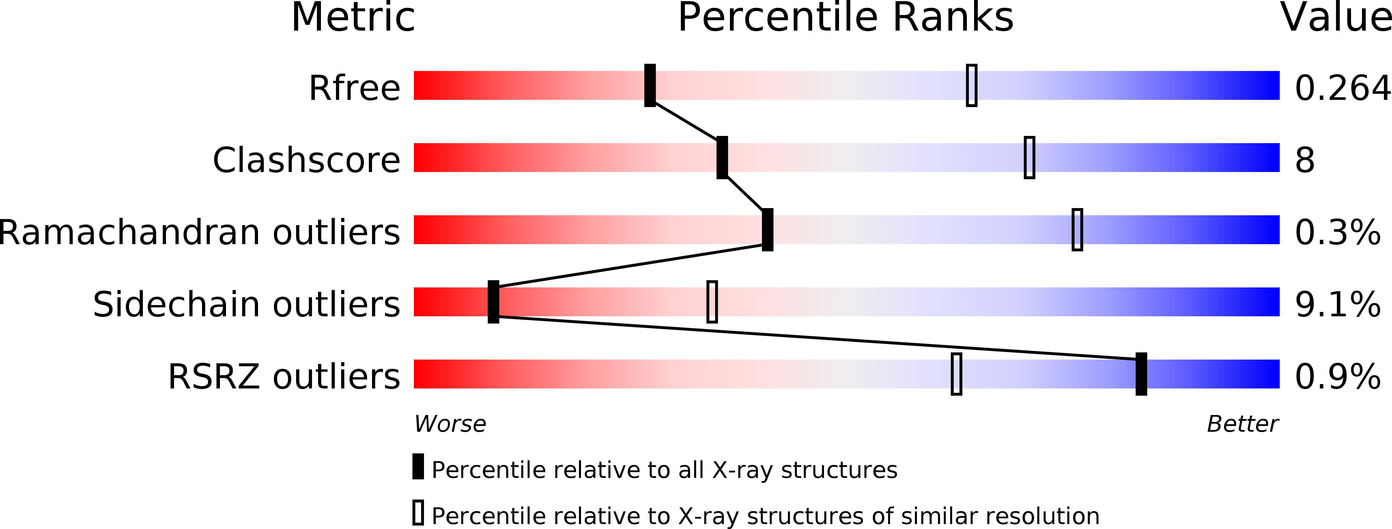
0.26
R-Value Work:
0.22
R-Value Observed:
0.22
Space Group:
P 62 2 2
Macromolecular Entities
Structures with similar UniProt ID
Protein Blast
Polymer Type:polypeptide(L)
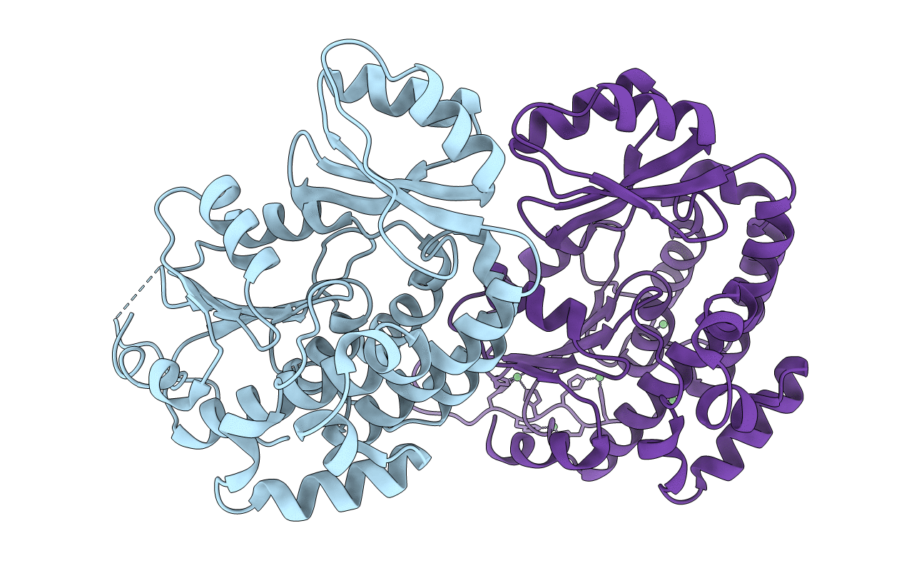
Molecule:Malonyl CoA-acyl carrier protein transacylase
Chain IDs:A, B
Chain Length:322
Number of Molecules:2
Biological Source:Mycobacterium tuberculosis
Ligand Molecules
Primary Citation
Structure of Mycobacterium tuberculosis mtFabD, a malonyl-CoA:acyl carrier protein transacylase (MCAT).
Acta Crystallogr.,Sect.F 63 831 835 (2007)
PMID: 17909282 DOI: 10.1107/S1744309107042455

Abstact

Mycobacteria display a unique and unusual cell-wall architecture, central to which is the membrane-proximal mycolyl-arabinogalactan-peptidoglycan core (mAGP). The biosynthesis of mycolic acids, which form the outermost layer of the mAGP core, involves malonyl-CoA:acyl carrier protein transacylase (MCAT). This essential enzyme catalyses the transfer of malonyl from coenzyme A to acyl carrier protein AcpM, thus feeding these two-carbon units into the chain-elongation cycle of the type II fatty-acid synthase. The crystal structure of M. tuberculosis mtFabD, the mycobacterial MCAT, has been determined to 3.0 A resolution by multi-wavelength anomalous dispersion. Phasing was facilitated by Ni2+ ions bound to the 20-residue N-terminal affinity tag, which packed between the two independent copies of mtFabD.

Legend

Protein

Chemical

Disease

Primary Citation of related structures