2QIA image
Deposition Date 2007-07-03
Release Date 2007-10-02
Last Version Date 2023-08-30
Entry Detail
PDB ID:
2QIA
Keywords:
Title:
Structural basis for the acyl chain selectivity and mechanism of UDP-N-acetylglucosamine Acyltransferase
Biological Source:
Source Organism:
Host Organism:
Method Details:
Experimental Method:
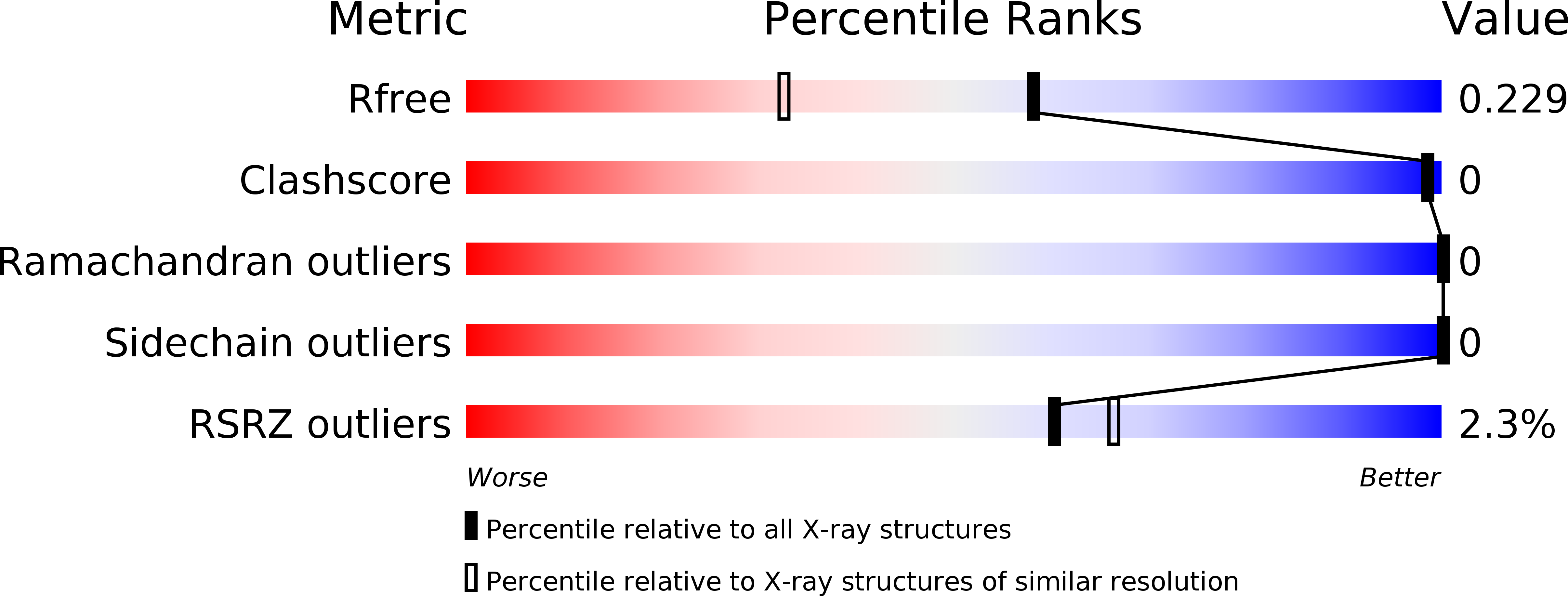
Resolution:
1.74 Å
R-Value Free:
0.23
R-Value Work:
0.18
R-Value Observed:
0.18
Space Group:
P 21 3
Macromolecular Entities
Structures with similar UniProt ID
Protein Blast
Polymer Type:polypeptide(L)
Molecule:UDP-N-acetylglucosamine acyltransferase
Gene (Uniprot):lpxA
Chain IDs:A
Chain Length:262
Number of Molecules:1
Biological Source:Escherichia coli K12
Ligand Molecules
Primary Citation
Structural basis for the acyl chain selectivity and mechanism of UDP-N-acetylglucosamine acyltransferase
Proc.Natl.Acad.Sci.Usa 104 13543 13550 (2007)
PMID: 17698807 DOI: 10.1073/pnas.0705833104

Abstact

UDP-N-acetylglucosamine (UDP-GlcNAc) acyltransferase (LpxA) catalyzes the first step of lipid A biosynthesis, the reversible transfer of the R-3-hydroxyacyl chain from R-3-hydroxyacyl acyl carrier protein to the glucosamine 3-OH group of UDP-GlcNAc. Escherichia coli LpxA is highly selective for R-3-hydroxymyristate. The crystal structure of the E. coli LpxA homotrimer, determined previously in the absence of lipid substrates or products, revealed that LpxA contains an unusual, left-handed parallel beta-helix fold. We have now solved the crystal structures of E. coli LpxA with the bound product UDP-3-O-(R-3-hydroxymyristoyl)-GlcNAc at a resolution of 1.74 A and with bound UDP-3-O-(R-3-hydroxydecanoyl)-GlcNAc at 1.85 A. The structures of these complexes are consistent with the catalytic mechanism deduced by mutagenesis and with a recent 3.0-A structure of LpxA with bound UDP-GlcNAc. Our structures show how LpxA selects for 14-carbon R-3-hydroxyacyl chains and reveal two modes of UDP binding.

Legend

Protein

Chemical

Disease

Primary Citation of related structures