2QGI image
Deposition Date 2007-06-29
Release Date 2008-01-22
Last Version Date 2023-08-30
Entry Detail
PDB ID:
2QGI
Keywords:
Title:
The UDP complex structure of the sixth gene product of the F1-ATPase operon of Rhodobacter blasticus
Biological Source:
Source Organism:
Host Organism:
Method Details:
Experimental Method:
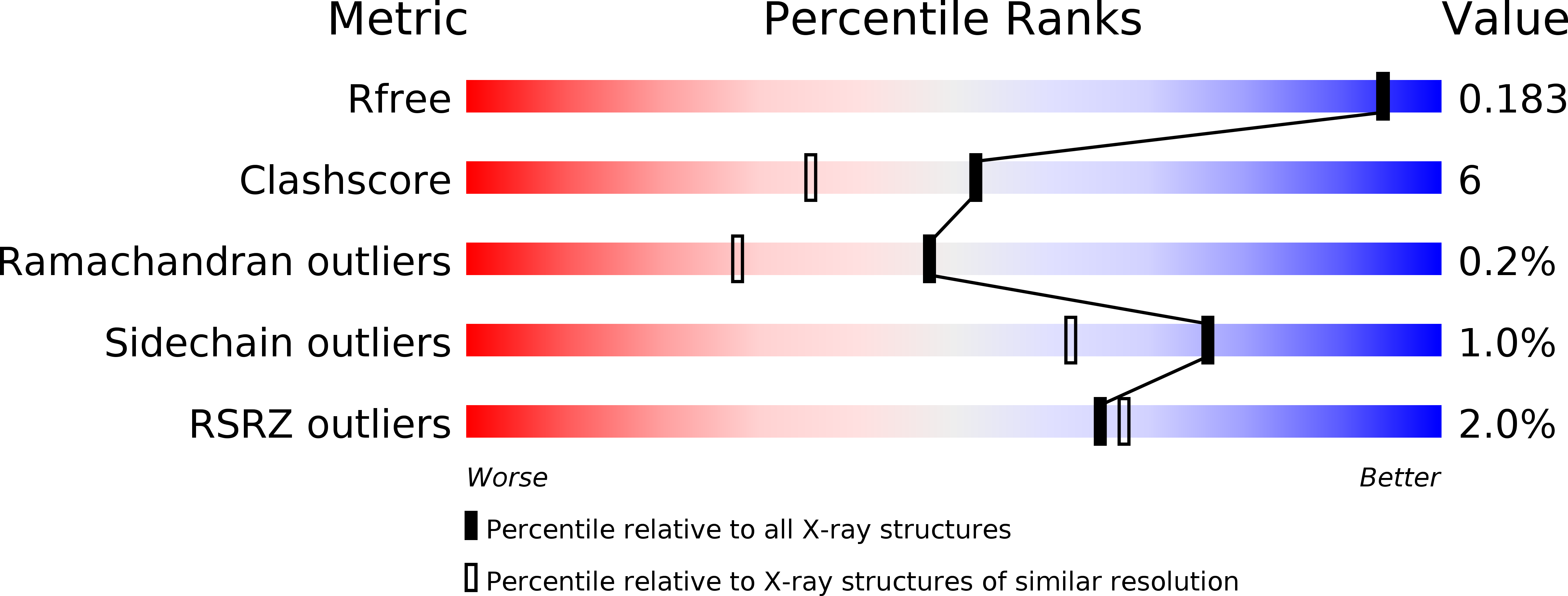
Resolution:
1.65 Å
R-Value Free:
0.18
R-Value Work:
0.15
R-Value Observed:
0.16
Space Group:
P 21 21 2
Macromolecular Entities
Structures with similar UniProt ID
Protein Blast
Polymer Type:polypeptide(L)
Molecule:ATP synthase subunits region ORF 6
Chain IDs:A, B
Chain Length:248
Number of Molecules:2
Biological Source:Rhodobacter blasticus
Primary Citation
Crystal structure of a protein, structurally related to glycosyltransferases, encoded in the Rhodobacter blasticus atp operon.
Biochim.Biophys.Acta 1784 379 384 (2008)
PMID: 18067873 DOI: 10.1016/j.bbapap.2007.11.005

Abstact

The F1-ATP synthase atp operon in the proteobacterium Rhodobacter blasticus contains six open reading frames, encoding six hypothetical proteins. Five of these subunits, in the stoichiometry (alphabeta)3gamma delta epsilon make up the catalytic F1-ATP synthase complex similarly in bacteria, chloroplasts and mitochondria. The sixth gene of the R. blasticus atp operon, urf6, shows very little sequence homology to any protein of known structure or function. The gene has previously been cloned, the product (called majastridin) has been heterologously expressed in Escherichia coli, and purified to high homogeneity [M. Brosché, I. Kalbina, M. Arnfelt, G. Benito, B.G. Karlsson, A. Strid, Occurrence, overexpression and partial purification of the protein (majastridin) corresponding to the URF6 gene of the Rhodobacter blasticus atp operon, Eur. J. Biochem. 255 (1998) 87-92]. We have solved the X-ray crystal structure and refined a model of majastridin to atomic resolution. Here we present the crystal structures of apo-majastridin and the complex of majastridin with Mn2+ and UDP and show that it has extensive structural similarity to glycosyltransferases (EC 2.4). This is the first structure determined from a new group of distantly related bacterial proteins of at least six members. They share the identical amino acids that bind Mn2+ and a triplet of amino acids in the putative sugar-binding site.

Legend

Protein

Chemical

Disease

Primary Citation of related structures
Feedback Form
Name
Email
Institute
Feedback