Deposition Date
2007-06-27
Release Date
2008-03-04
Last Version Date
2024-02-21
Entry Detail
PDB ID:
2QFJ
Keywords:
Title:
Crystal Structure of First Two RRM Domains of FIR Bound to ssDNA from a Portion of FUSE
Biological Source:
Source Organism(s):
Homo sapiens (Taxon ID: 9606)
Expression System(s):
Method Details:
Experimental Method:
Resolution:
2.10 Å
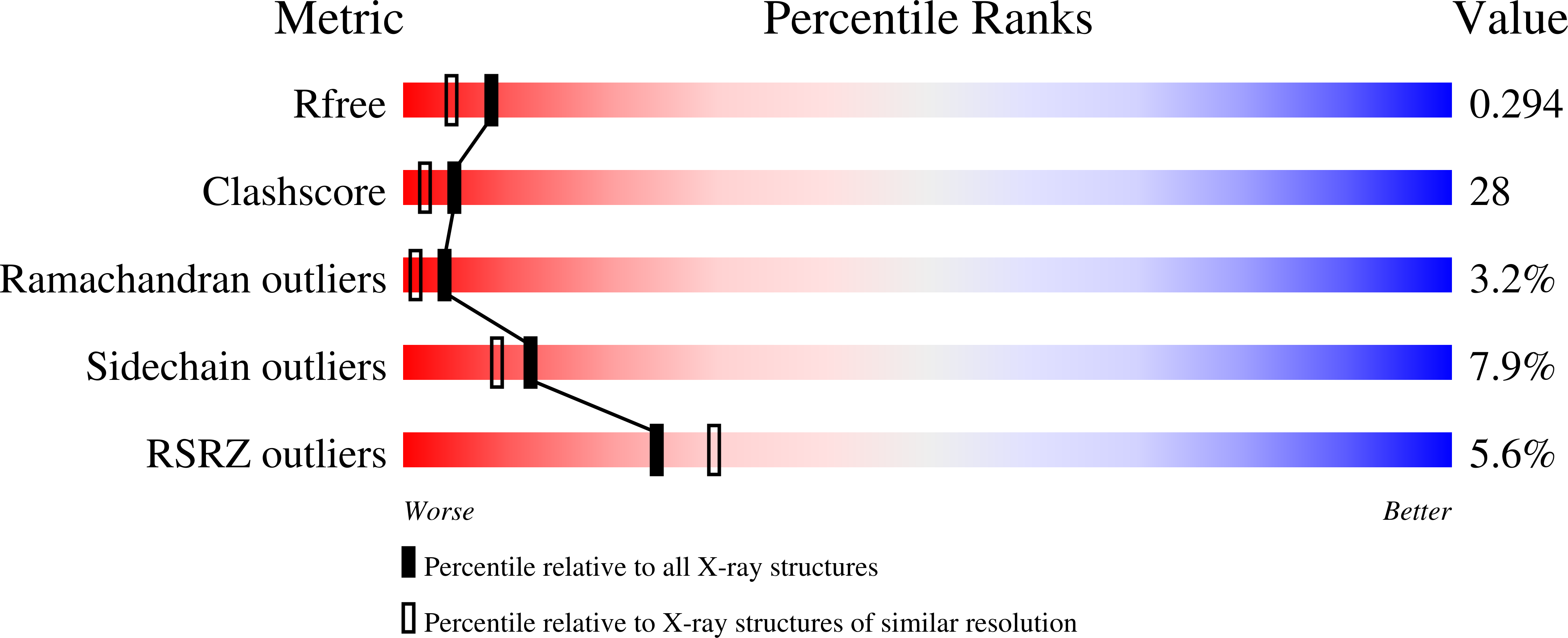
R-Value Free:
0.29
R-Value Work:
0.25
R-Value Observed:
0.25
Space Group:
P 31