2QFD image
Deposition Date 2007-06-27
Release Date 2008-02-12
Last Version Date 2024-10-30
Entry Detail
PDB ID:
2QFD
Keywords:
Title:
Crystal structure of the regulatory domain of human RIG-I with bound Hg
Biological Source:
Source Organism:
Homo sapiens (Taxon ID: 9606)
Host Organism:
Method Details:
Experimental Method:
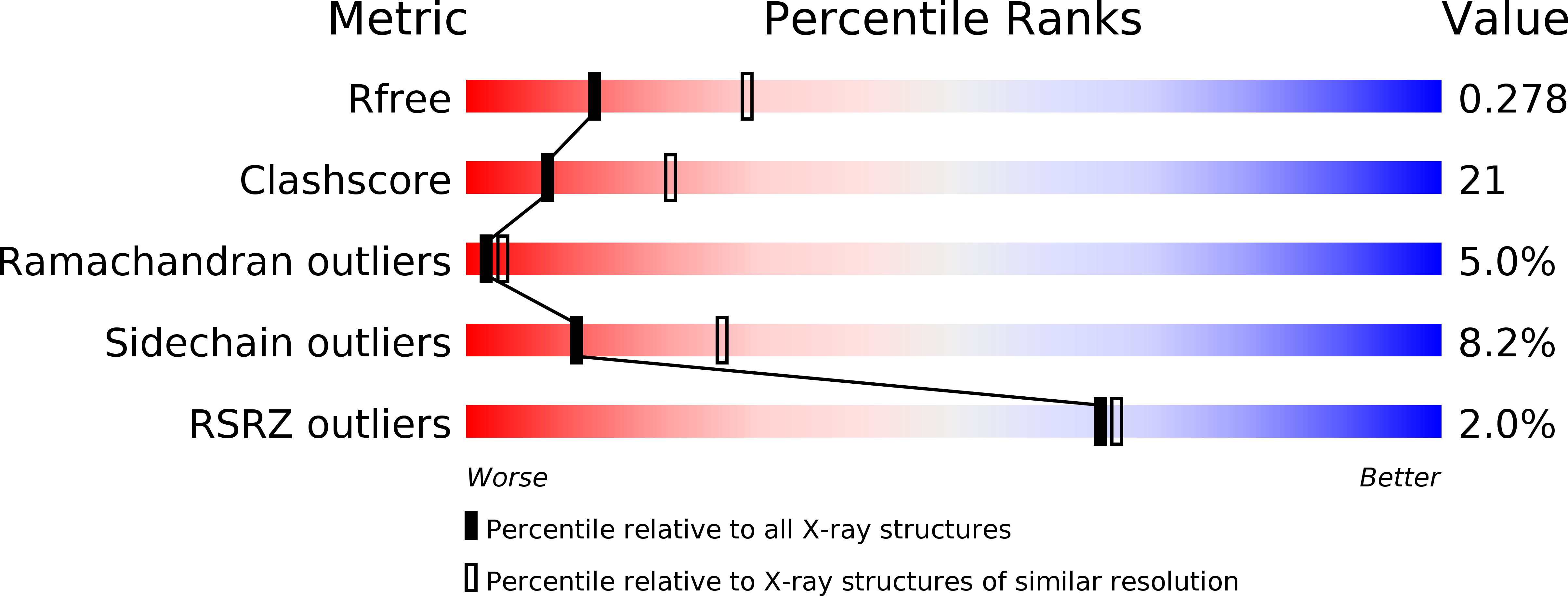
Resolution:
2.70 Å
R-Value Free:
0.27
R-Value Work:
0.24
R-Value Observed:
0.25
Space Group:
P 1 21 1
Macromolecular Entities
Polymer Type:polypeptide(L)
Molecule:Probable ATP-dependent RNA helicase DDX58
Gene (Uniprot):RIGI
Chain IDs:A, B, C, D, E, F, G, H, I, J
Chain Length:145
Number of Molecules:10
Biological Source:Homo sapiens
Ligand Molecules
Primary Citation
The C-Terminal Regulatory Domain Is the RNA 5'-Triphosphate Sensor of RIG-I.
Mol.Cell 29 169 179 (2008)
PMID: 18243112 DOI: 10.1016/j.molcel.2007.10.032

Abstact

The ATPase RIG-I senses viral RNAs that contain 5'-triphosphates in the cytoplasm. It initiates a signaling cascade that activates innate immune response by interferon and cytokine production, providing essential antiviral protection for the host. The mode of RNA 5'-triphosphate sensing by RIG-I remains elusive. We show that the C-terminal regulatory domain RD of RIG-I binds viral RNA in a 5'-triphosphate-dependent manner and activates the RIG-I ATPase by RNA-dependent dimerization. The crystal structure of RD reveals a zinc-binding domain that is structurally related to GDP/GTP exchange factors of Rab-like GTPases. The zinc coordination site is essential for RIG-I signaling and is also conserved in MDA5 and LGP2, suggesting related RD domains in all three enzymes. Structure-guided mutagenesis identifies a positively charged groove as likely 5'-triphosphate-binding site of RIG-I. This groove is distinct in MDA5 and LGP2, raising the possibility that RD confers ligand specificity.

Legend

Protein

Chemical

Disease

Primary Citation of related structures