2QDY image
Deposition Date 2007-06-22
Release Date 2008-05-13
Last Version Date 2024-10-30
Entry Detail
PDB ID:
2QDY
Keywords:
Title:
Crystal Structure of Fe-type NHase from Rhodococcus erythropolis AJ270
Biological Source:
Source Organism:
Method Details:
Experimental Method:
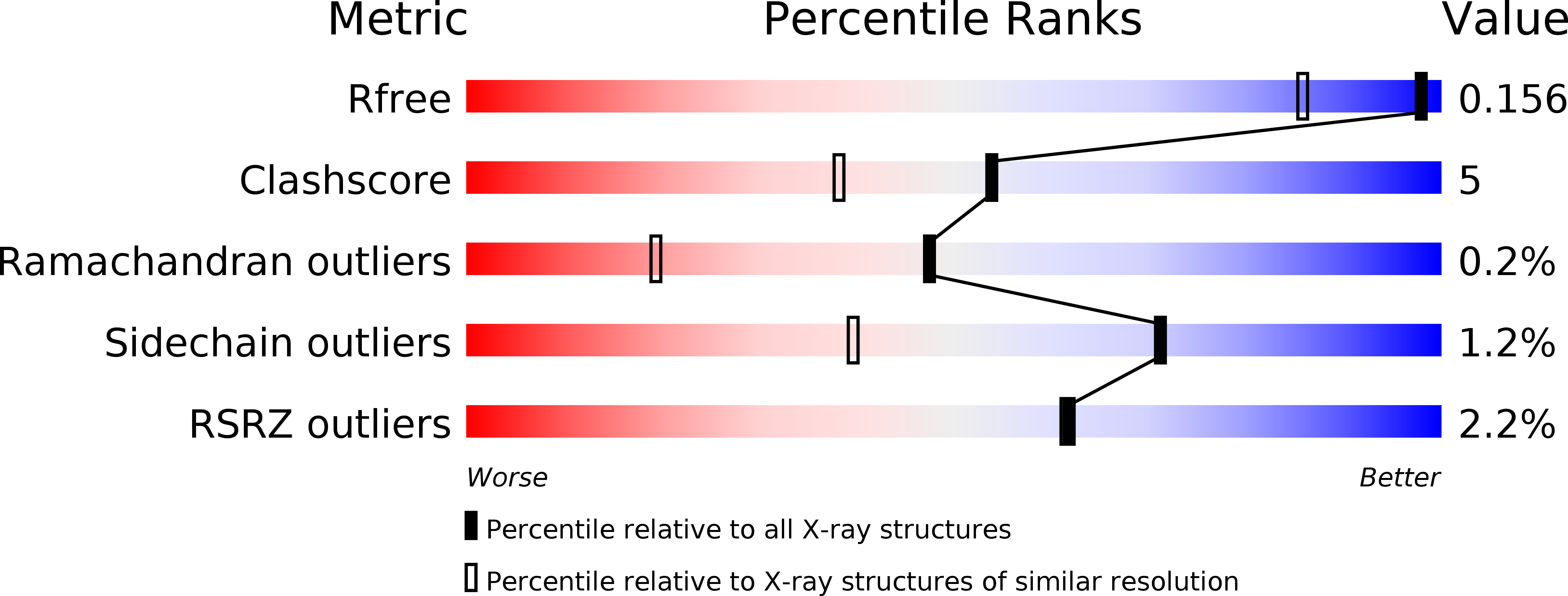
Resolution:
1.30 Å
R-Value Free:
0.15
R-Value Work:
0.12
R-Value Observed:
0.13
Space Group:
C 1 2 1
Macromolecular Entities
Polymer Type:polypeptide(L)
Molecule:Nitrile hydratase subunit alpha
Gene (Uniprot):nthA
Chain IDs:A
Chain Length:207
Number of Molecules:1
Biological Source:Rhodococcus erythropolis
Polymer Type:polypeptide(L)
Molecule:Nitrile hydratase subunit beta
Gene (Uniprot):nthB
Chain IDs:B
Chain Length:212
Number of Molecules:1
Biological Source:Rhodococcus erythropolis
Modified Residue
Compound ID Chain ID Parent Comp ID Details 2D Image
CSD A CYS 3-SULFINOALANINE
Primary Citation
High resolution X-ray molecular structure of the nitrile hydratase from Rhodococcus erythropolis AJ270 reveals posttranslational oxidation of two cysteines into sulfinic acids and a novel biocatalytic nitrile hydration mechanism
Biochem.Biophys.Res.Commun. 362 319 324 (2007)
PMID: 17716629 DOI: 10.1016/j.bbrc.2007.07.184

Abstact

The crystal structure of Fe-type nitrile hydratase from Rhodococcus erythropolis AJ270 was determined at 1.3A resolution. The two cysteine residues (alphaCys(112) and alphaCys(114)) equatorially coordinated to the ferric ion were post-translationally modified to cysteine sulfinic acids. A glutamine residue (alphaGln(90)) in the active center gave double conformations. Based on the interactions among the enzyme, substrate and water molecules, a new mechanism of biocatalysis of nitrile hydratase was proposed, in which the water molecule activated by the glutamine residue performed as the nucleophile to attack on the nitrile which was simultaneously interacted by another water molecule coordinated to the ferric ion.

Legend

Protein

Chemical

Disease

Primary Citation of related structures