Deposition Date
2007-06-14
Release Date
2008-07-08
Last Version Date
2024-11-06
Entry Detail
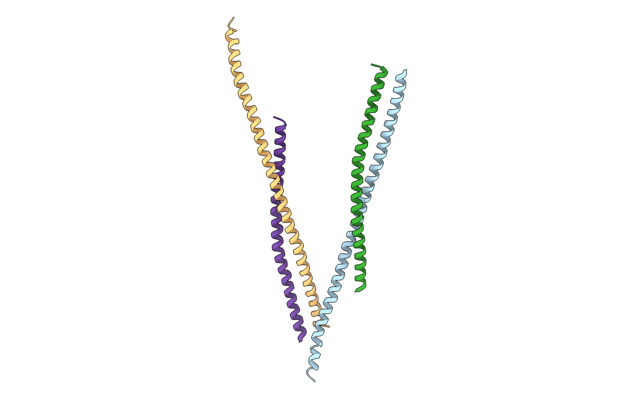
PDB ID:
2QA7
Keywords:
Title:
Crystal structure of Huntingtin-interacting protein 1 (HIP1) coiled-coil domain with a basic surface suitable for HIP-protein interactor (HIPPI)
Biological Source:
Source Organism(s):
Homo sapiens (Taxon ID: 9606)
Expression System(s):
Method Details:
Experimental Method:
Resolution:
2.80 Å
R-Value Free:
0.32
R-Value Work:
0.26
R-Value Observed:
0.32
Space Group:
P 43