2Q91 image
Entry Detail
PDB ID:
2Q91
Title:
Structure of the Ca2+-Bound Activated Form of the S100A4 Metastasis Factor
Biological Source:
Source Organism:
Host Organism:
PDB Version:
Deposition Date:
2007-06-12
Release Date:
2008-02-26
Method Details:
Experimental Method:
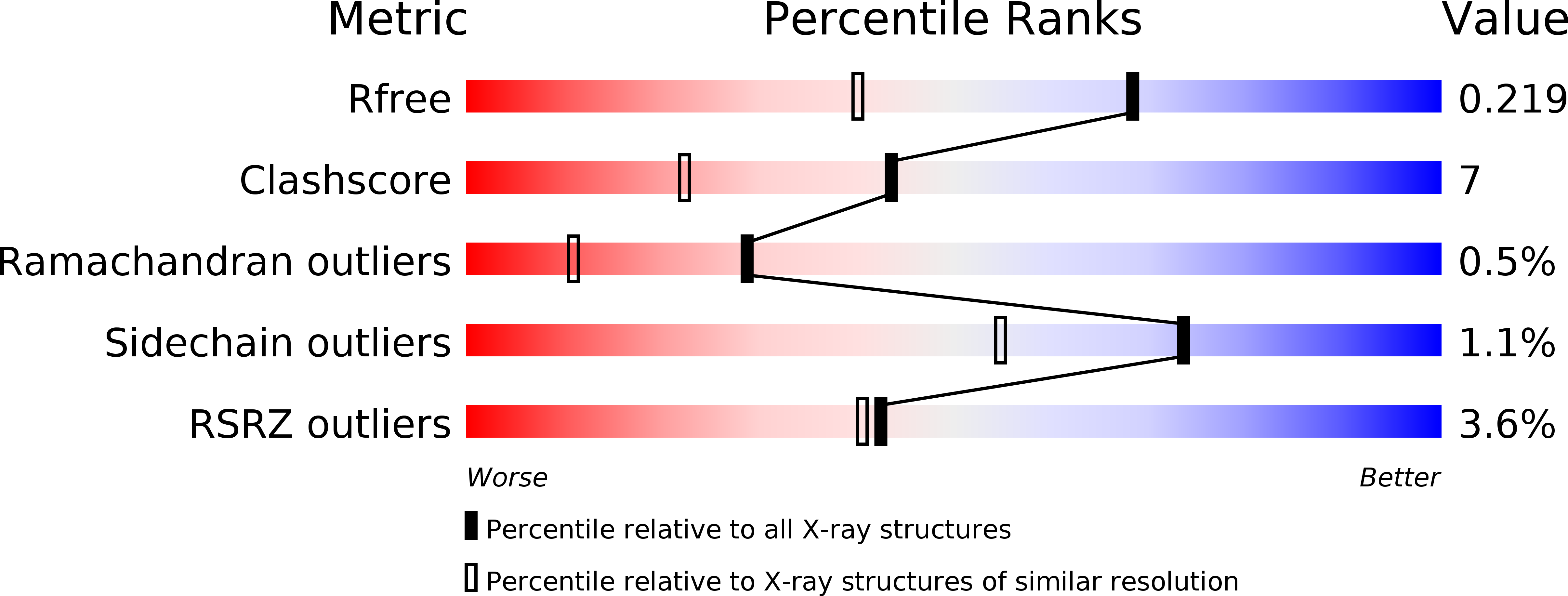
Resolution:
1.63 Å
R-Value Free:
0.21
R-Value Work:
0.18
R-Value Observed:
0.18
Space Group:
P 32 2 1
Macromolecular Entities
Polymer Type:polypeptide(L)
Description:S100A4 Metastasis Factor
Chain IDs:A, B
Chain Length:101
Number of Molecules:2
Biological Source:Homo sapiens
Ligand Molecules
Primary Citation
Structure of Ca(2+)-Bound S100A4 and Its Interaction with Peptides Derived from Nonmuscle Myosin-IIA.
Biochemistry 47 5111 5126 (2008)
PMID: 18410126 DOI: 10.1021/bi702537s

Abstact

S100A4, also known as mts1, is a member of the S100 family of Ca2+-binding proteins that is directly involved in tumor invasion and metastasis via interactions with specific protein targets, including nonmuscle myosin-IIA (MIIA). Human S100A4 binds two Ca2+ ions with the typical EF-hand exhibiting an affinity that is nearly 1 order of magnitude tighter than that of the pseudo-EF-hand. To examine how Ca2+ modifies the overall organization and structure of the protein, we determined the 1.7 A crystal structure of the human Ca2+-S100A4. Ca2+ binding induces a large reorientation of helix 3 in the typical EF-hand. This reorganization exposes a hydrophobic cleft that is comprised of residues from the hinge region,helix 3, and helix 4, which afford specific target recognition and binding. The Ca2+-dependent conformational change is required for S100A4 to bind peptide sequences derived from the C-terminal portion of the MIIA rod with submicromolar affinity. In addition, the level of binding of Ca2+ to both EF-hands increases by 1 order of magnitude in the presence of MIIA. NMR spectroscopy studies demonstrate that following titration with a MIIA peptide, the largest chemical shift perturbations and exchange broadening effects occur for residues in the hydrophobic pocket of Ca2+-S100A4. Most of these residues are not exposed in apo-S100A4 and explain the Ca2+ dependence of formation of theS100A4-MIIA complex. These studies provide the foundation for understanding S100A4 target recognition and may support the development of reagents that interfere with S100A4 function.

Legend

Protein

Chemical

Disease

Primary Citation of related structures