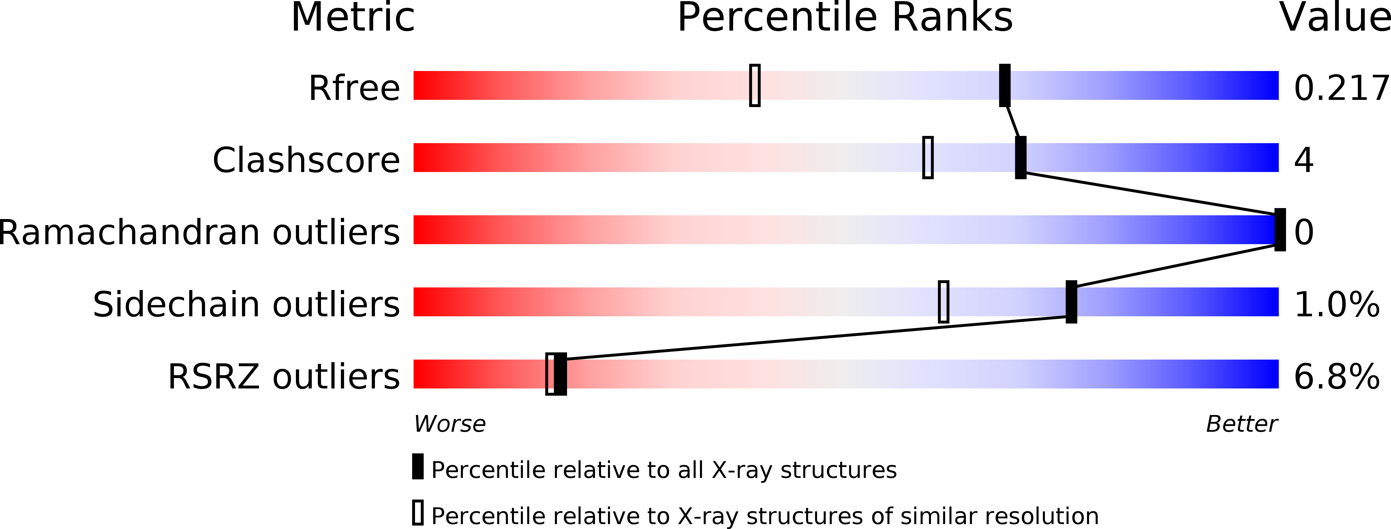
2Q8K image
Deposition Date 2007-06-11
Release Date 2007-09-25
Last Version Date 2023-08-30
Entry Detail
PDB ID:
2Q8K
Keywords:
Title:
The crystal structure of Ebp1
Biological Source:
Source Organism:
Homo sapiens (Taxon ID: 9606)
Method Details:
Experimental Method:
Resolution:
1.60 Å
R-Value Free:
0.21
R-Value Work:
0.18
R-Value Observed:
0.18
Space Group:
P 41 21 2
Macromolecular Entities
Structures with similar UniProt ID
Protein Blast
Polymer Type:polypeptide(L)
Molecule:Proliferation-associated protein 2G4
Gene (Uniprot):PA2G4
Chain IDs:A
Chain Length:401
Number of Molecules:1
Biological Source:Homo sapiens
Primary Citation
The crystal structure of Ebp1 reveals a methionine aminopeptidase fold as binding platform for multiple interactions.
Febs Lett. 581 4450 4454 (2007)
PMID: 17765895 DOI: 10.1016/j.febslet.2007.08.024

Abstact

The ErbB-3 receptor binding protein (Ebp1) is a member of the proliferation-associated 2G4 (PA2G4) family implicated in regulation of cell growth and differentiation. Here, we report the crystal structure of the human Ebp1 at 1.6 A resolution. The protein has the conserved pita bread fold of methionine aminopeptidases, but without the characteristic enzymatic activity. Moreover, Ebp1 is known to interact with a number of proteins and RNAs involved in either transcription regulation or translation control. The structure provides insights in how Ebp1 discriminates between its different interaction partners.

Legend

Protein

Chemical

Disease

Primary Citation of related structures