
Deposition Date
2007-06-06
Release Date
2007-09-18
Last Version Date
2024-02-21
Entry Detail
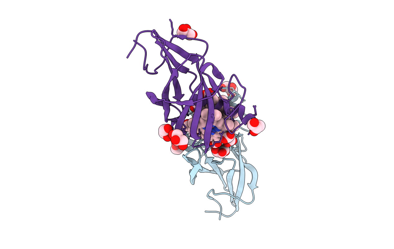
PDB ID:
2Q7A
Keywords:
Title:
Crystal structure of the cell surface heme transfer protein Shp
Biological Source:
Source Organism(s):
Streptococcus pyogenes (Taxon ID: 1314)
Expression System(s):
Method Details:
Experimental Method:
Resolution:
2.10 Å
R-Value Free:
0.22
R-Value Work:
0.16
R-Value Observed:
0.16
Space Group:
P 65
