2Q5S image
Entry Detail
PDB ID:
2Q5S
Title:
Crystal Structure of PPARgamma bound to partial agonist nTZDpa
Biological Source:
Source Organism:
Host Organism:
PDB Version:
Deposition Date:
2007-06-01
Release Date:
2007-10-23
Method Details:
Experimental Method:
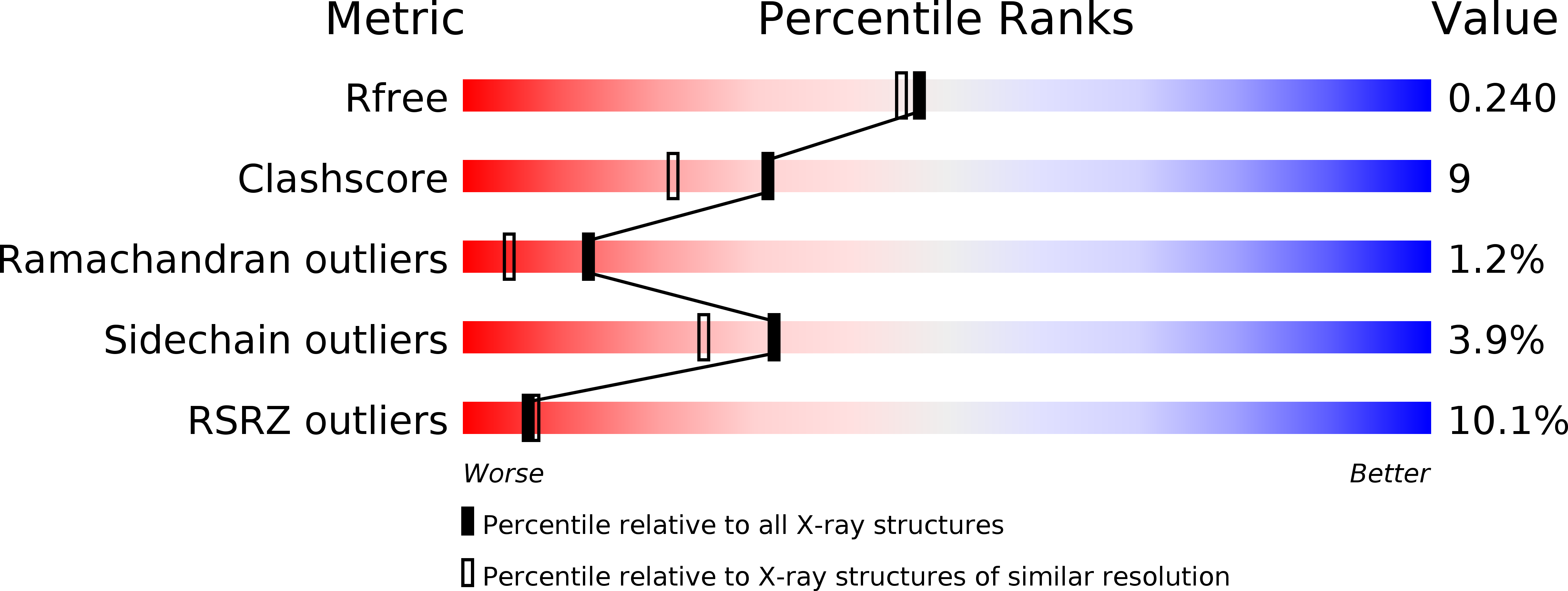
Resolution:
2.05 Å
R-Value Free:
0.24
R-Value Work:
0.19
R-Value Observed:
0.19
Space Group:
C 1 2 1
Macromolecular Entities
Polymer Type:polypeptide(L)
Description:Peroxisome Proliferator-Activated Receptor gamma
Chain IDs:A, B
Chain Length:274
Number of Molecules:2
Biological Source:Homo sapiens
Ligand Molecules
Primary Citation
Partial Agonists Activate PPARgamma Using a Helix 12 Independent Mechanism
Structure 15 1258 1271 (2007)
PMID: 17937915 DOI: 10.1016/j.str.2007.07.014

Abstact

Binding to helix 12 of the ligand-binding domain of PPARgamma is required for full agonist activity. Previously, the degree of stabilization of the activation function 2 (AF-2) surface was thought to correlate with the degree of agonism and transactivation. To examine this mechanism, we probed structural dynamics of PPARgamma with agonists that induced graded transcriptional responses. Here we present crystal structures and amide H/D exchange (HDX) kinetics for six of these complexes. Amide HDX revealed each ligand induced unique changes to the dynamics of the ligand-binding domain (LBD). Full agonists stabilized helix 12, whereas intermediate and partial agonists did not at all, and rather differentially stabilized other regions of the binding pocket. The gradient of PPARgamma transactivation cannot be accounted for solely through changes to the dynamics of AF-2. Thus, our understanding of allosteric signaling must be extended beyond the idea of a dynamic helix 12 acting as a molecular switch.

Legend

Protein

Chemical

Disease

Primary Citation of related structures