2Q31 image
Entry Detail
PDB ID:
2Q31
Title:
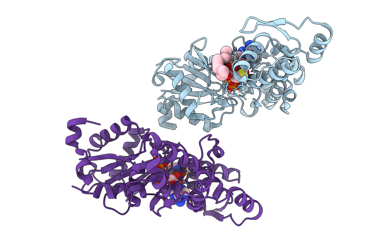
Actin Dimer Cross-linked Between Residues 41 and 374 and proteolytically cleaved by subtilisin between residues 47 and 48.
Biological Source:
Source Organism:
PDB Version:
Deposition Date:
2007-05-29
Release Date:
2007-06-05
Method Details:
Experimental Method:
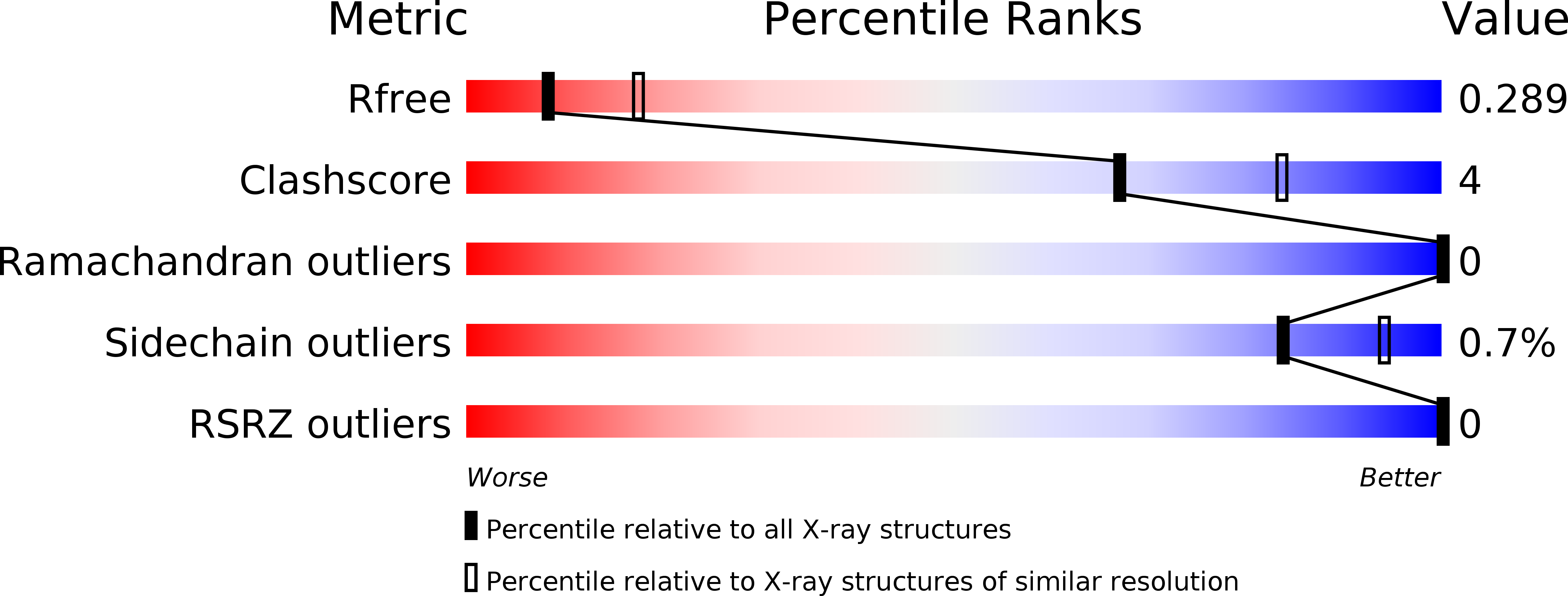
Resolution:
2.70 Å
R-Value Free:
0.28
R-Value Work:
0.24
R-Value Observed:
0.25
Space Group:
P 1 21 1
Macromolecular Entities
Polymer Type:polypeptide(L)
Description:Actin, alpha skeletal muscle
Chain IDs:A, B
Chain Length:375
Number of Molecules:2
Biological Source:Oryctolagus cuniculus
Primary Citation
Multiple crystal structures of actin dimers and their implications for interactions in the actin filament.
Acta Crystallogr.,Sect.D 64 454 465 (2008)
PMID: 18391412 DOI: 10.1107/S0907444908003351

Abstact

The structure of actin in its monomeric form is known at high resolution, while the structure of filamentous F-actin is only understood at considerably lower resolution. Knowing precisely how the monomers of actin fit together would lead to a deeper understanding of the dynamic behavior of the actin filament. Here, a series of crystal structures of actin dimers are reported which were prepared by cross-linking in either the longitudinal or the lateral direction in the filament state. Laterally cross-linked dimers, comprised of monomers belonging to different protofilaments, are found to adopt configurations in crystals that are not related to the native structure of filamentous actin. In contrast, multiple structures of longitudinal dimers consistently reveal the same interface between monomers within a single protofilament. The reappearance of the same longitudinal interface in multiple crystal structures adds weight to arguments that the interface visualized is similar to that in actin filaments. Highly conserved atomic interactions involving residues 199-205 and 287-291 are highlighted.

Legend

Protein

Chemical

Disease

Primary Citation of related structures