2Q2B image
Deposition Date 2007-05-27
Release Date 2007-07-17
Last Version Date 2023-08-30
Entry Detail
PDB ID:
2Q2B
Keywords:
Title:
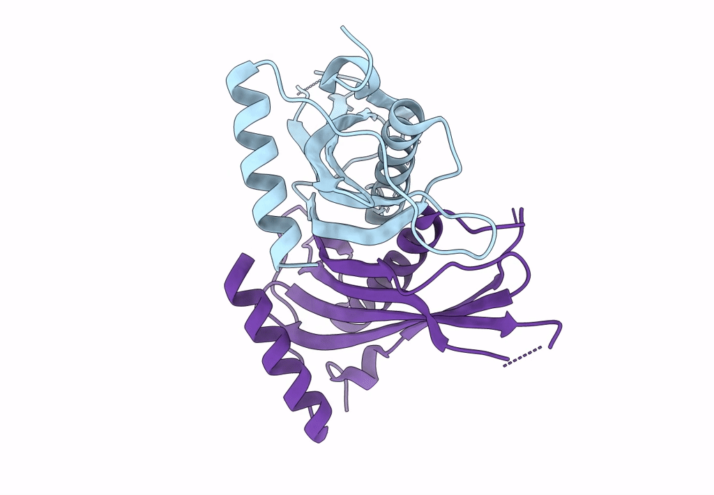
Crystal structure of the C-terminal domain of mouse acyl-CoA thioesterase 7
Biological Source:
Source Organism:
Mus musculus (Taxon ID: 10090)
Host Organism:
Method Details:
Experimental Method:
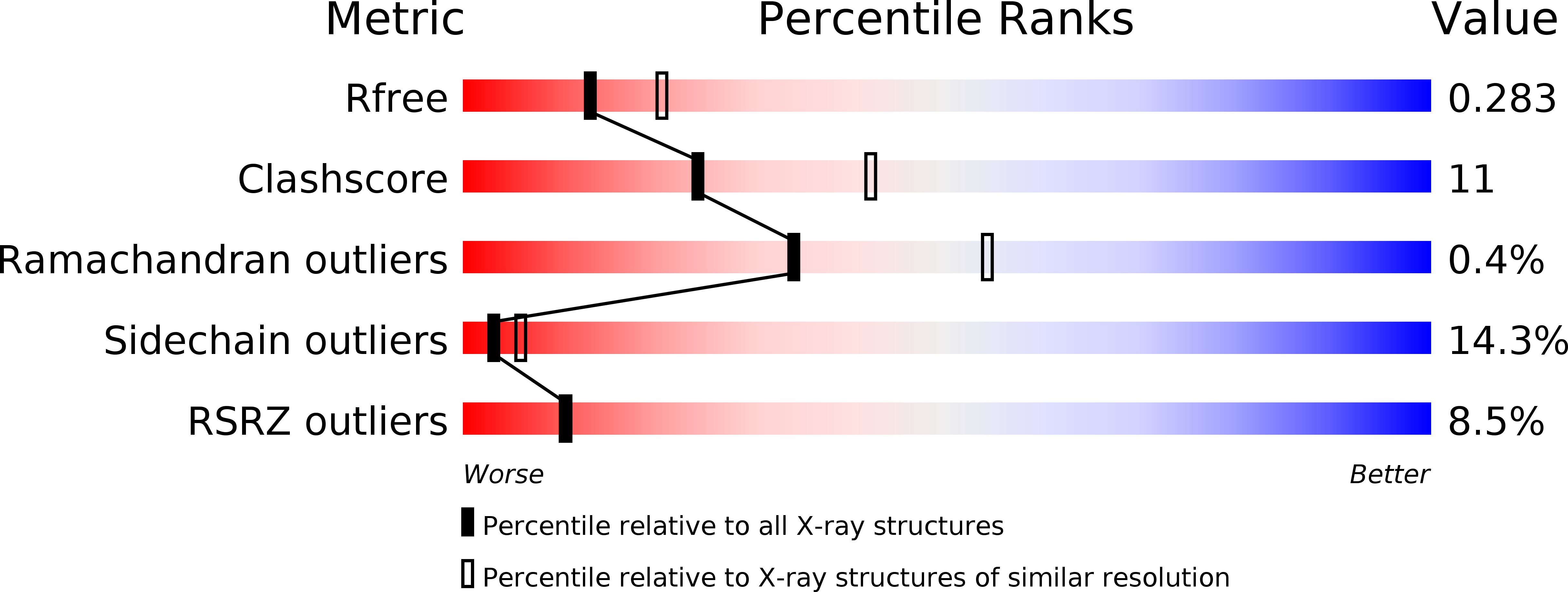
Resolution:
2.50 Å
R-Value Free:
0.28
R-Value Work:
0.22
R-Value Observed:
0.22
Space Group:
H 3 2
Macromolecular Entities
Structures with similar UniProt ID
Protein Blast
Polymer Type:polypeptide(L)
Molecule:Cytosolic acyl coenzyme A thioester hydrolase
Gene (Uniprot):Acot7
Chain IDs:A, B
Chain Length:179
Number of Molecules:2
Biological Source:Mus musculus
Primary Citation
Structural basis for recruitment of tandem hotdog domains in acyl-CoA thioesterase 7 and its role in inflammation.
Proc.Natl.Acad.Sci.Usa 104 10382 10387 (2007)
PMID: 17563367 DOI: 10.1073/pnas.0700974104

Abstact

Acyl-CoA thioesterases (Acots) catalyze the hydrolysis of fatty acyl-CoA to free fatty acid and CoA and thereby regulate lipid metabolism and cellular signaling. We present a comprehensive structural and functional characterization of mouse acyl-CoA thioesterase 7 (Acot7). Whereas prokaryotic homologues possess a single thioesterase domain, mammalian Acot7 contains a pair of domains in tandem. We determined the crystal structures of both the N- and C-terminal domains of the mouse enzyme, and inferred the structure of the full-length enzyme using a combination of chemical cross-linking, mass spectrometry, and molecular modeling. The quaternary arrangement in Acot7 features a trimer of hotdog fold dimers. Both domains of Acot7 are required for activity, but only one of two possible active sites in the dimer is functional. Asn-24 and Asp-213 (from N- and C-domains, respectively) were identified as the catalytic residues through site-directed mutagenesis. An enzyme with higher activity than wild-type Acot7 was obtained by mutating the residues in the nonfunctional active site. Recombinant Acot7 was shown to have the highest activity toward arachidonoyl-CoA, suggesting a function in eicosanoid metabolism. In line with the proposal, Acot7 was shown to be highly expressed in macrophages and up-regulated by lipopolysaccharide. Overexpression of Acot7 in a macrophage cell line modified the production of prostaglandins D2 and E2. Together, the results link the molecular and cellular functions of Acot7 and identify the enzyme as a candidate drug target in inflammatory disease.

Legend

Protein

Chemical

Disease

Primary Citation of related structures
Feedback Form
Name
Email
Institute
Feedback