2Q29 image
Deposition Date 2007-05-26
Release Date 2008-06-03
Last Version Date 2024-02-21
Entry Detail
PDB ID:
2Q29
Keywords:
Title:
Crystal structure of oxalyl-coA decarboxylase from Escherichia coli in complex with acetyl coenzyme A
Biological Source:
Source Organism:
Escherichia coli (Taxon ID: )
Host Organism:
Method Details:
Experimental Method:
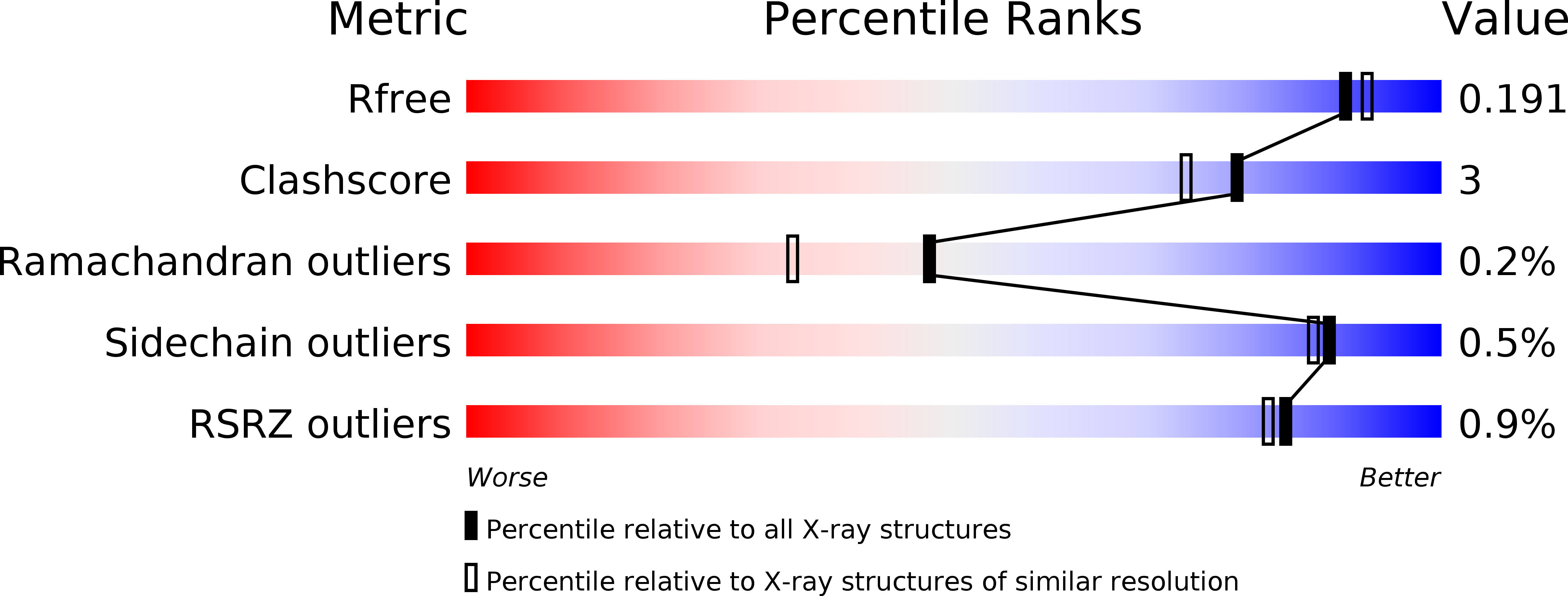
Resolution:
1.82 Å
R-Value Free:
0.19
R-Value Work:
0.17
R-Value Observed:
0.17
Space Group:
C 2 2 21
Macromolecular Entities
Polymer Type:polypeptide(L)
Molecule:oxalyl-CoA decarboxylase
Gene (Uniprot):oxc
Chain IDs:A, B
Chain Length:564
Number of Molecules:2
Biological Source:Escherichia coli
Primary Citation
New insights into structure-function relationships of oxalyl CoA decarboxylase from Escherichia coli.
Febs J. 277 2628 2640 (2010)
PMID: 20553497 DOI: 10.1111/j.1742-464X.2010.07673.x

Abstact

The gene yfdU from Escherichia coli encodes a putative oxalyl coenzyme A decarboxylase, a thiamine diphosphate-dependent enzyme that is potentially involved in the degradation of oxalate. The enzyme has been purified to homogeneity. The kinetic constants for conversion of the substrate oxalyl coenzyme A by the enzyme in the absence and presence of the inhibitor coenzyme A, as well as in the absence and presence of the activator adenosine 5'-diphosphate, were determined using a novel continuous optical assay. The effects of these ligands on the solution and crystal structure of the enzyme were studied using small-angle X-ray scattering and X-ray crystal diffraction. Analyses of the obtained crystal structures of the enzyme in complex with the cofactor thiamine diphosphate, the activator adenosine 5'-diphosphate and the inhibitor acetyl coenzyme A, as well as the corresponding solution scattering patterns, allow comparison of the oligomer structures of the enzyme complexes under various experimental conditions, and provide insights into the architecture of substrate and effector binding sites.

Legend

Protein

Chemical

Disease

Primary Citation of related structures