Deposition Date
2007-05-22
Release Date
2007-07-03
Last Version Date
2024-10-30
Entry Detail
PDB ID:
2Q0L
Keywords:
Title:
Helicobacter pylori thioredoxin reductase reduced by sodium dithionite in complex with NADP+
Biological Source:
Source Organism(s):
Helicobacter pylori (Taxon ID: 85962)
Expression System(s):
Method Details:
Experimental Method:
Resolution:
1.45 Å
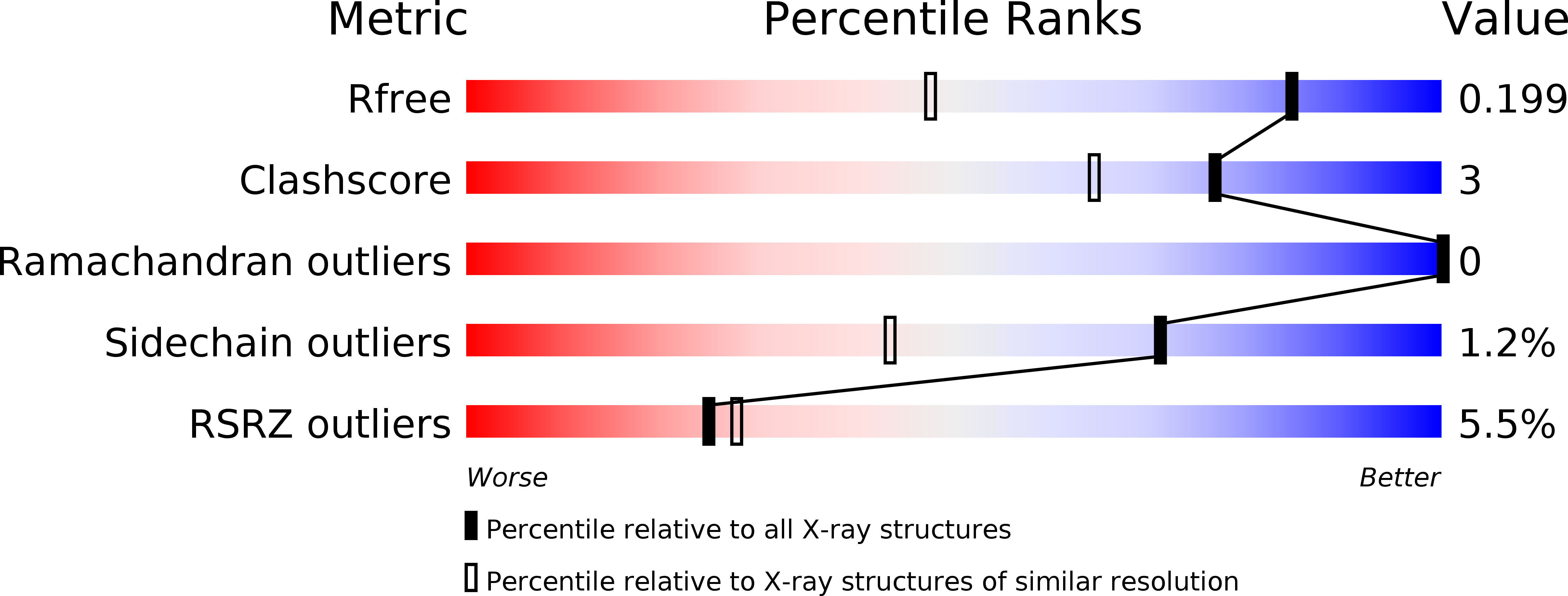
R-Value Free:
0.19
R-Value Work:
0.16
R-Value Observed:
0.16
Space Group:
P 1 21 1