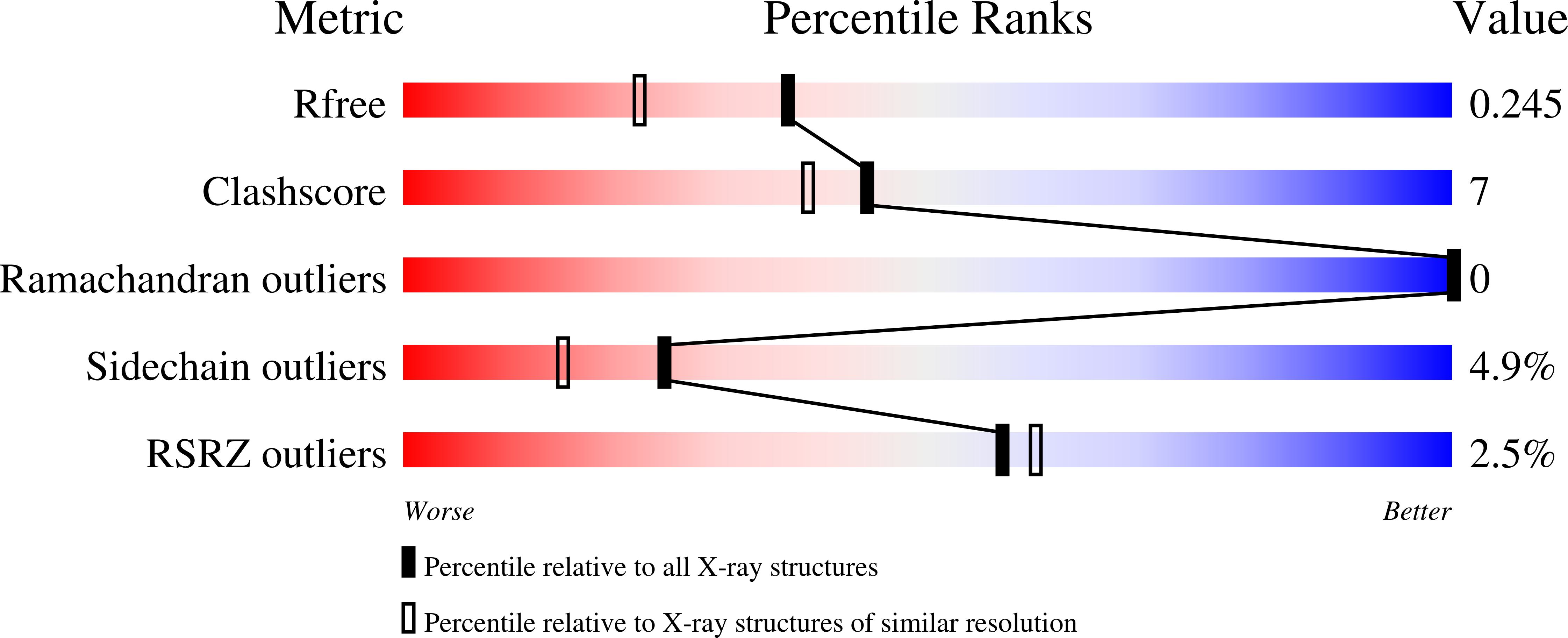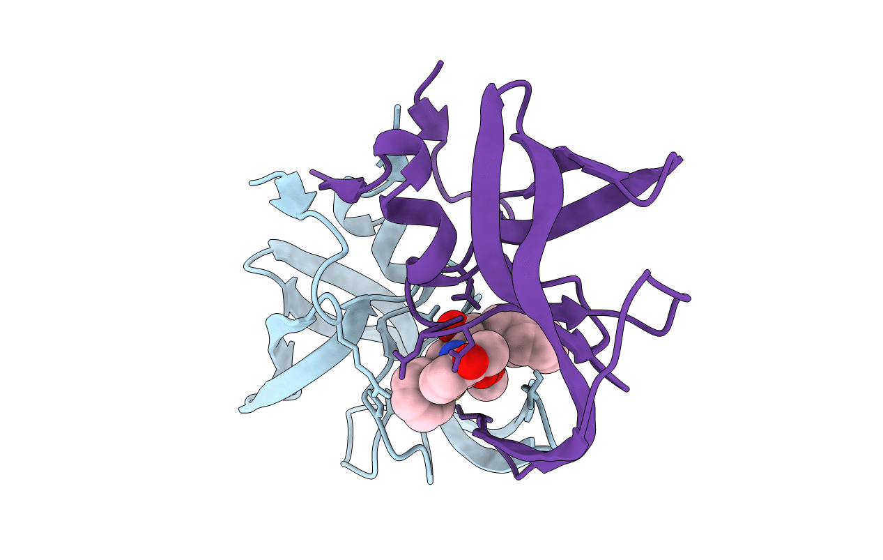
2PYM image
Deposition Date 2007-05-16
Release Date 2008-02-26
Last Version Date 2023-08-30
Entry Detail
PDB ID:
2PYM
Keywords:
Title:
HIV-1 PR mutant in complex with nelfinavir
Biological Source:
Source Organism:
Method Details:
Experimental Method:
Resolution:
1.90 Å
R-Value Free:
0.24
R-Value Work:
0.19
R-Value Observed:
0.19
Space Group:
P 61
Macromolecular Entities
Polymer Type:polypeptide(L)
Molecule:PROTEASE RETROPEPSIN
Gene (Uniprot):gag-pol
Mutations:D30N, N88D
Chain IDs:A, B
Chain Length:99
Number of Molecules:2
Biological Source:Human immunodeficiency virus 1
Ligand Molecules
Primary Citation
Molecular analysis of the HIV-1 resistance development: enzymatic activities, crystal structures, and thermodynamics of nelfinavir-resistant HIV protease mutants
J.Mol.Biol. 374 1005 1016 (2007)
PMID: 17977555 DOI: 10.1016/j.jmb.2007.09.083

Abstact

Human immunodeficiency virus (HIV) encodes an aspartic protease (PR) that cleaves viral polyproteins into mature proteins, thus leading to the formation of infectious particles. Protease inhibitors (PIs) are successful virostatics. However, their efficiency is compromised by antiviral resistance. In the PR sequence of viral variants resistant to the PI nelfinavir, the mutations D30N and L90M appear frequently. However, these two mutations are seldom found together in vivo, suggesting that there are two alternative evolutionary pathways leading to nelfinavir resistance. Here we analyze the proteolytic activities, X-ray structures, and thermodynamics of inhibitor binding to HIV-1 PRs harboring the D30N and L90M mutations alone and in combination with other compensatory mutations. Vitality values obtained for recombinant mutant proteases and selected PR inhibitors confirm the crucial role of mutations in positions 30 and 90 for nelfinavir resistance. The combination of the D30N and L90M mutations significantly increases the enzyme vitality in the presence of nelfinavir, without a dramatic decrease in the catalytic efficiency of the recombinant enzyme. Crystal structures, molecular dynamics simulations, and calorimetric data for four mutants (D30N, D30N/A71V, D30N/N88D, and D30N/L90M) were used to augment our kinetic data. Calorimetric analysis revealed that the entropic contribution to the mutant PR/nelfinavir interaction is less favorable than the entropic contribution to the binding of nelfinavir by wild-type PR. This finding is supported by the structural data and simulations; nelfinavir binds most strongly to the wild-type protease, which has the lowest number of protein-ligand hydrogen bonds and whose structure exhibits the greatest degree of fluctuation upon inhibitor binding.

Legend

Protein

Chemical

Disease

Primary Citation of related structures