2PWL image
Deposition Date 2007-05-11
Release Date 2007-09-25
Last Version Date 2023-08-30
Entry Detail
PDB ID:
2PWL
Keywords:
Title:
Crystal Structure of FGF Receptor 2 (FGFR2) Kinase Domain Harboring the Pathogenic N549H Mutation Responsible for Crouzon Syndrome.
Biological Source:
Source Organism:
Homo sapiens (Taxon ID: 9606)
Host Organism:
Method Details:
Experimental Method:
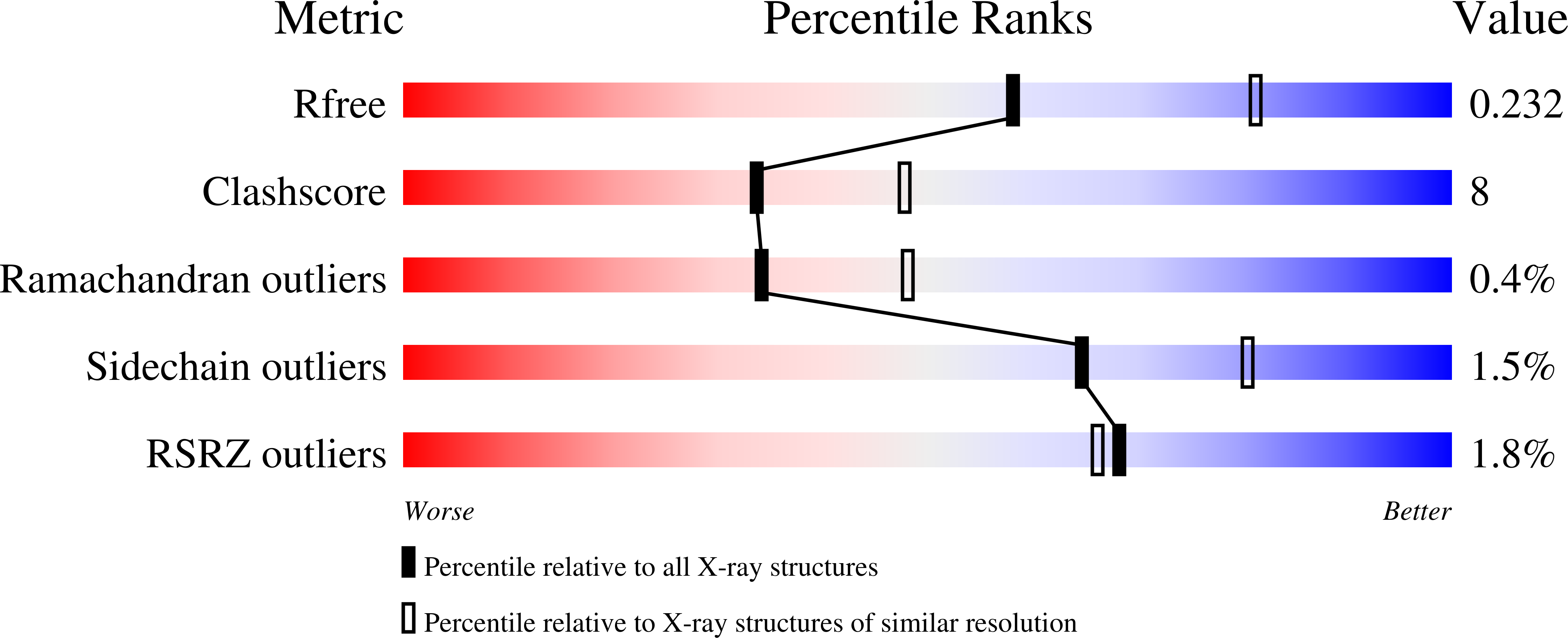
Resolution:
2.40 Å
R-Value Free:
0.23
R-Value Work:
0.20
Space Group:
P 21 21 2
Macromolecular Entities
Polymer Type:polypeptide(L)
Molecule:Fibroblast growth factor receptor 2
Gene (Uniprot):FGFR2
Mutations:C491A, N549H
Chain IDs:A, B
Chain Length:324
Number of Molecules:2
Biological Source:Homo sapiens
Primary Citation
A molecular brake in the kinase hinge region regulates the activity of receptor tyrosine kinases.
Mol.Cell 27 717 730 (2007)
PMID: 17803937 DOI: 10.1016/j.molcel.2007.06.028

Abstact

Activating mutations in the tyrosine kinase domain of receptor tyrosine kinases (RTKs) cause cancer and skeletal disorders. Comparison of the crystal structures of unphosphorylated and phosphorylated wild-type FGFR2 kinase domains with those of seven unphosphorylated pathogenic mutants reveals an autoinhibitory "molecular brake" mediated by a triad of residues in the kinase hinge region of all FGFRs. Structural analysis shows that many other RTKs, including PDGFRs, VEGFRs, KIT, CSF1R, FLT3, TEK, and TIE, are also subject to regulation by this brake. Pathogenic mutations activate FGFRs and other RTKs by disengaging the brake either directly or indirectly.

Legend

Protein

Chemical

Disease

Primary Citation of related structures