2PVF image
Deposition Date 2007-05-09
Release Date 2007-09-25
Last Version Date 2024-10-30
Entry Detail
PDB ID:
2PVF
Keywords:
Title:
Crystal Structure of Tyrosine Phosphorylated Activated FGF Receptor 2 (FGFR2) Kinase Domain in Complex with ATP Analog and Substrate Peptide
Biological Source:
Source Organism:
Homo sapiens (Taxon ID: 9606)
(Taxon ID: )
Host Organism:
Method Details:
Experimental Method:
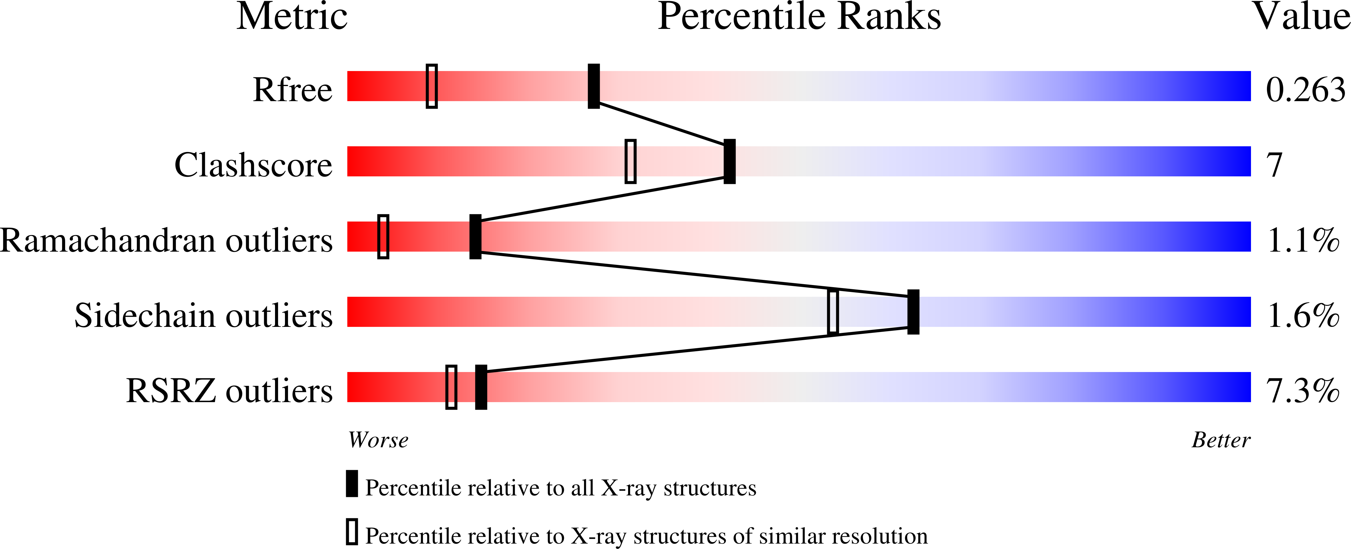
Resolution:
1.80 Å
R-Value Free:
0.26
R-Value Work:
0.25
Space Group:
P 21 21 21
Macromolecular Entities
Polymer Type:polypeptide(L)
Molecule:Fibroblast growth factor receptor 2
Gene (Uniprot):FGFR2
Mutations:C491A
Chain IDs:A
Chain Length:334
Number of Molecules:1
Biological Source:Homo sapiens
Polymer Type:polypeptide(L)
Molecule:Fibroblast growth factor receptor 2
Gene (Uniprot):FGFR2
Chain IDs:B
Chain Length:15
Number of Molecules:1
Biological Source:
Modified Residue
Compound ID Chain ID Parent Comp ID Details 2D Image
PTR A TYR O-PHOSPHOTYROSINE
Primary Citation
A molecular brake in the kinase hinge region regulates the activity of receptor tyrosine kinases.
Mol.Cell 27 717 730 (2007)
PMID: 17803937 DOI: 10.1016/j.molcel.2007.06.028

Abstact

Activating mutations in the tyrosine kinase domain of receptor tyrosine kinases (RTKs) cause cancer and skeletal disorders. Comparison of the crystal structures of unphosphorylated and phosphorylated wild-type FGFR2 kinase domains with those of seven unphosphorylated pathogenic mutants reveals an autoinhibitory "molecular brake" mediated by a triad of residues in the kinase hinge region of all FGFRs. Structural analysis shows that many other RTKs, including PDGFRs, VEGFRs, KIT, CSF1R, FLT3, TEK, and TIE, are also subject to regulation by this brake. Pathogenic mutations activate FGFRs and other RTKs by disengaging the brake either directly or indirectly.

Legend

Protein

Chemical

Disease

Primary Citation of related structures