Deposition Date
2007-05-09
Release Date
2007-07-10
Last Version Date
2024-11-20
Entry Detail
PDB ID:
2PV9
Keywords:
Title:
Crystal structure of murine thrombin in complex with the extracellular fragment of murine PAR4
Biological Source:
Source Organism(s):
Mus musculus (Taxon ID: 10090)
Expression System(s):
Method Details:
Experimental Method:
Resolution:
3.50 Å
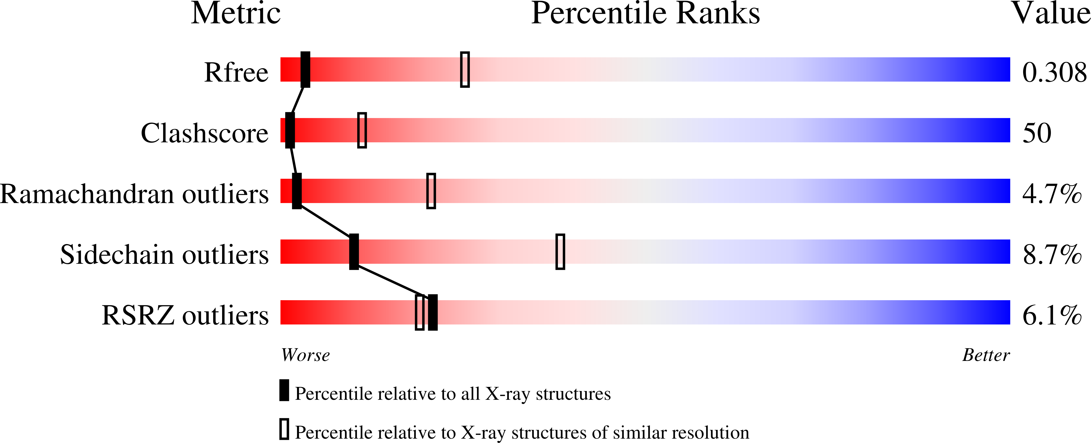
R-Value Free:
0.31
R-Value Work:
0.30
R-Value Observed:
0.30
Space Group:
P 65 2 2