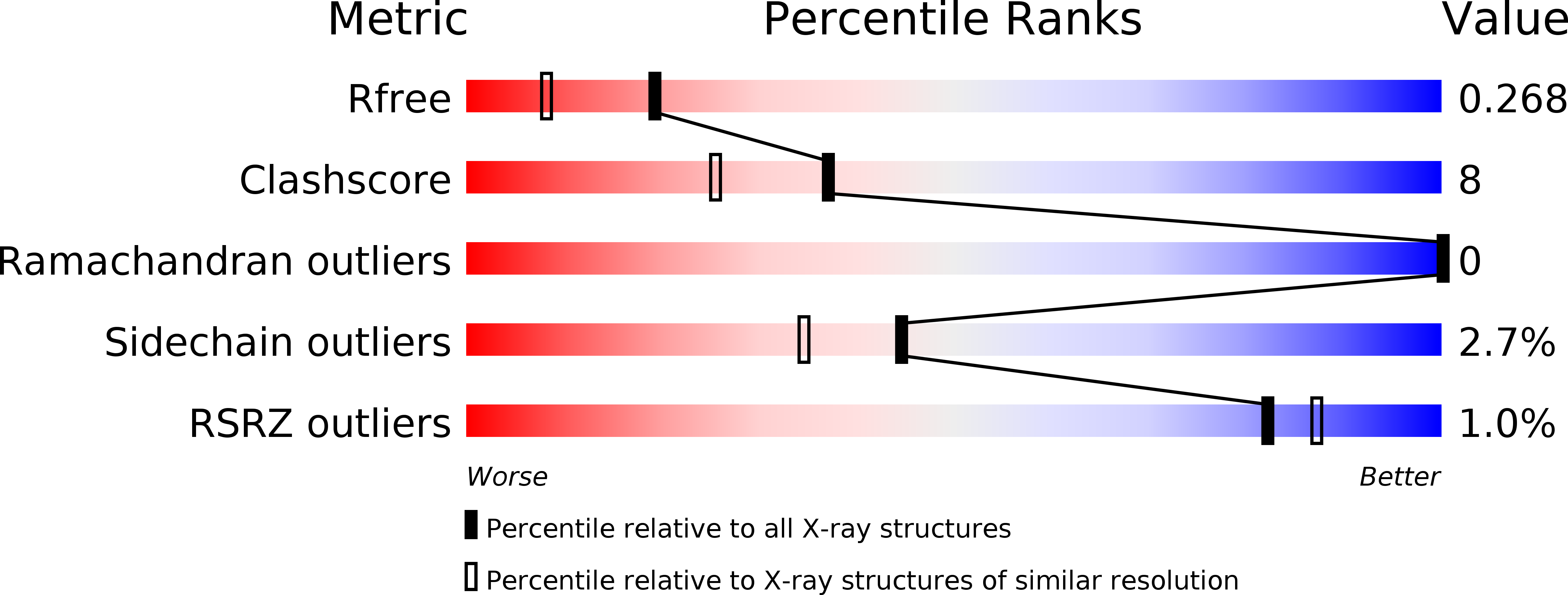
2PSP image
Deposition Date 1996-02-01
Release Date 1996-07-11
Last Version Date 2024-10-23
Entry Detail
PDB ID:
2PSP
Title:
Porcine pancreatic spasmolytic polypeptide
Biological Source:
Source Organism:
Sus scrofa (Taxon ID: )
Method Details:
Experimental Method:
Resolution:
1.95 Å
R-Value Free:
0.25
R-Value Work:
0.19
R-Value Observed:
0.19
Space Group:
P 31 2 1
Macromolecular Entities
Polymer Type:polypeptide(L)
Molecule:PORCINE PANCREATIC SPASMOLYTIC POLYPEPTIDE
Gene (Uniprot):TFF2
Chain IDs:A, B
Chain Length:106
Number of Molecules:2
Biological Source:Sus scrofa
Modified Residue
Compound ID Chain ID Parent Comp ID Details 2D Image
PCA A GLN PYROGLUTAMIC ACID
Primary Citation
Structure of porcine pancreatic spasmolytic polypeptide at 1.95 A resolution.
Acta Crystallogr.,Sect.D 52 730 737 (1996)
PMID: 15299636 DOI: 10.1107/S0907444996001345

Abstact

The structure of a trigonal crystal form of porcine pancreatic spasmolytic polypeptide (PSP) has been solved by molecular replacement and refined to 1.95 A resolution. Three heavy-atom derivatives were prepared, giving unbiased phase information, which was used in the model building of the protein molecules. The final conventional R value is 19.8% with the inclusion of 183 water molecules. PSP crystallizes as a dimer in space group P3(1)21 with a non-crystallographic twofold axis relating the monomers. The monomer consists of two very similar domains each composed of three loop regions. Two clefts are found in the monomer, one in each domain, that are proposed as possible substrate-binding sites. Important interactions have been identified in the proposed substrate-binding sites, where conserved water molecules probably mimic the hydrophilic positions of the substrate. The estimated cleft size is 9 x 9 x 12 A. Analysis of the charge distribution within the clefts, by an electrostatic potential calculation, shows the clefts to be essentially non-charged.

Legend

Protein

Chemical

Disease

Primary Citation of related structures