Deposition Date
1998-12-11
Release Date
1998-12-16
Last Version Date
2023-12-27
Entry Detail
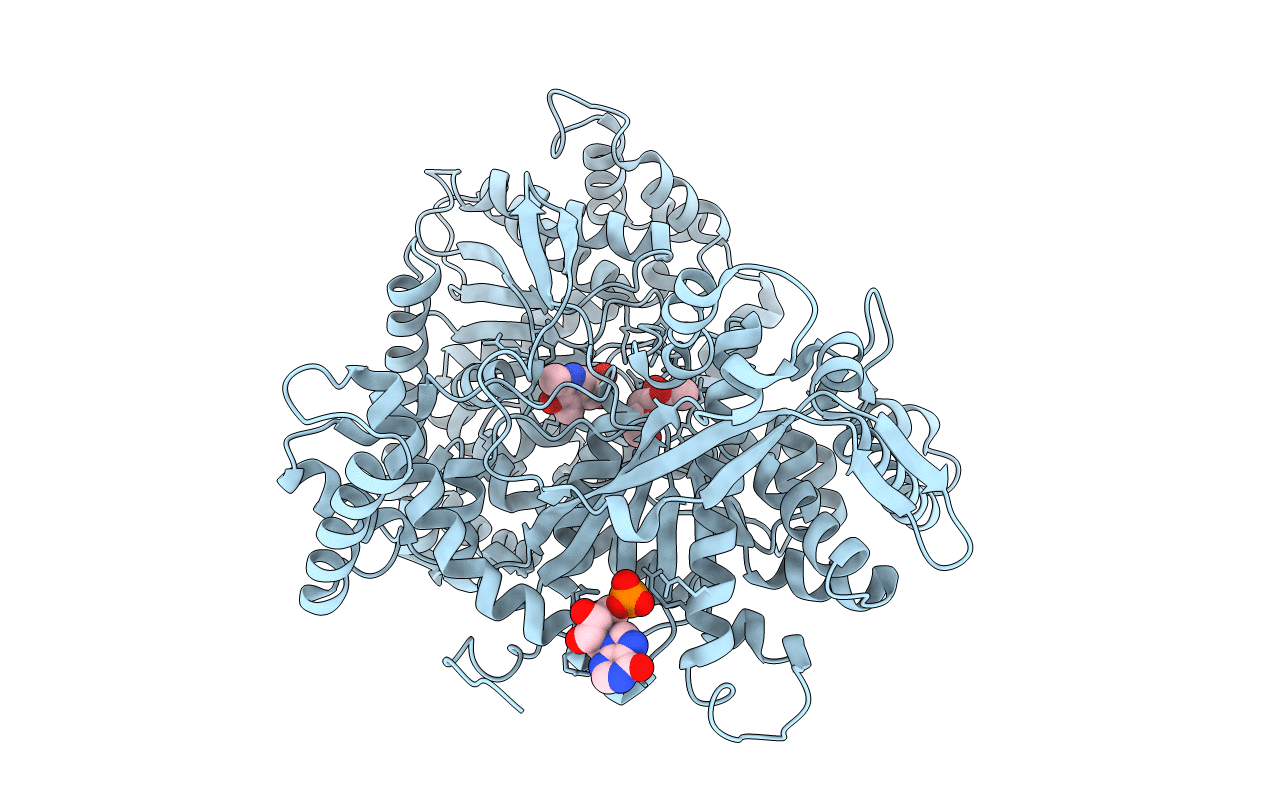
PDB ID:
2PRJ
Keywords:
Title:
Binding of N-acetyl-beta-D-glucopyranosylamine to Glycogen Phosphorylase B
Biological Source:
Source Organism(s):
Oryctolagus cuniculus (Taxon ID: 9986)
Method Details:
Experimental Method:
Resolution:
2.30 Å
R-Value Free:
0.23
R-Value Work:
0.18
R-Value Observed:
0.18
Space Group:
P 43 21 2