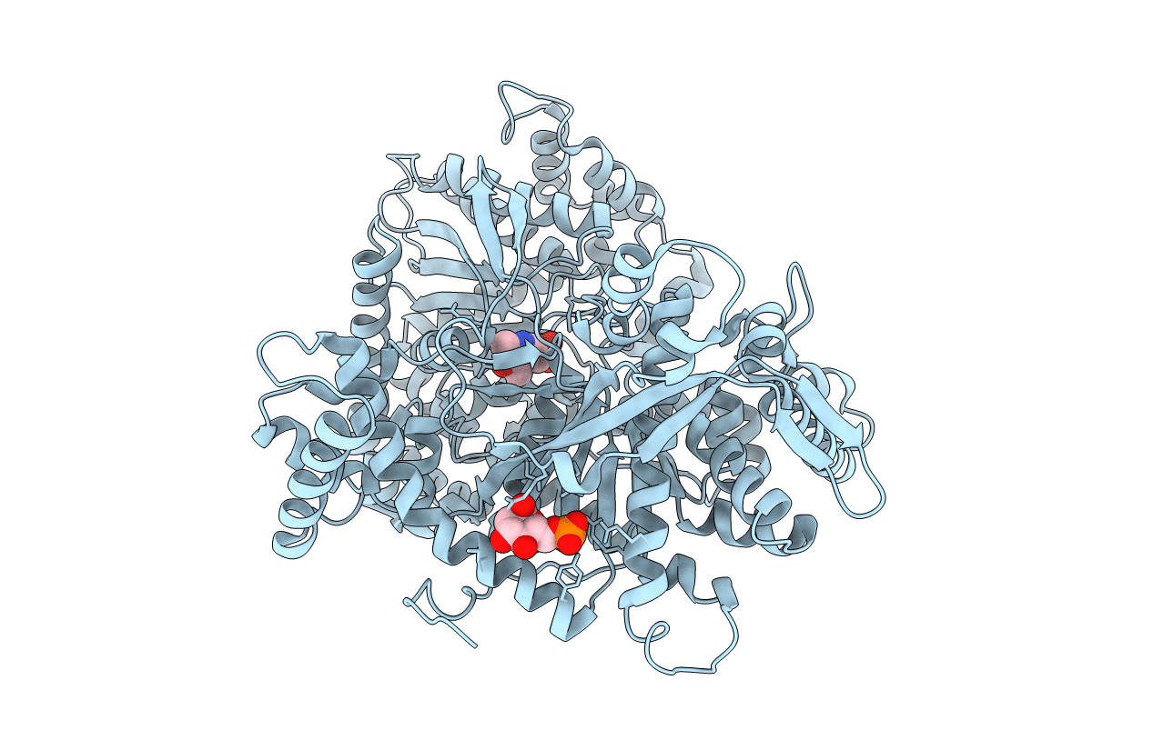
2PRI image
Deposition Date 1998-12-11
Release Date 1998-12-14
Last Version Date 2023-08-30
Entry Detail
PDB ID:
2PRI
Keywords:
Title:
BINDING OF 2-DEOXY-GLUCOSE-6-PHOSPHATE TO GLYCOGEN PHOSPHORYLASE B
Biological Source:
Source Organism:
Method Details:
Experimental Method:
Resolution:
2.30 Å
R-Value Free:
0.24
R-Value Work:
0.19
R-Value Observed:
0.19
Space Group:
P 43 21 2
Macromolecular Entities
Structures with similar UniProt ID
Protein Blast
Polymer Type:polypeptide(L)
Molecule:GLYCOGEN PHOSPHORYLASE B
Gene (Uniprot):PYGM
Chain IDs:A
Chain Length:842
Number of Molecules:1
Biological Source:Oryctolagus cuniculus
Primary Citation
The binding of 2-deoxy-D-glucose 6-phosphate to glycogen phosphorylase b: kinetic and crystallographic studies.
J.Mol.Biol. 254 900 917 (1995)
PMID: 7500360 DOI: 10.1006/jmbi.1995.0665

Abstact

Kinetic and crystallographic studies have characterized the effect of 2-deoxy-glucose 6-phosphate on the catalytic and structural properties of glycogen phosphorylase b. Previous work on the binding of glucose 6-phosphate, a potent physiological inhibitor of the enzyme, to T state phosphorylase b in the crystal showed that the inhibitor binds at the allosteric site and induces substantial conformational changes that affect the subunit-subunit interface. The hydrogen-bond from the O-2 hydroxyl of glucose 6-phosphate to the main-chain oxygen of Val40' represents the only hydrogen bond from the sugar to the other subunit, and this interaction appears important for promoting a more "tensed" structure than native T state phosphorylase b. 2-Deoxy-glucose 6-phosphate acts competitively with both the activator AMP and the substrate glucose 1-phosphate, with Ki values of 0.53 mM and 1.23 mM, respectively. The binding of 2-deoxy-glucose 6-phosphate to T state glycogen phosphorylase b in the crystal, has been investigated and the complex phosphorylase b: 2-deoxy-glucose 6-phosphate has been refined to give a crystallographic R factor of 17.3%, for data between 8 A and 2.3 A. 2-Deoxy-glucose 6-phosphate binds at the allosteric site as the a anomer and adopts a different conformation compared to glucose 6-phosphate. The two conformations differ by 160 degrees in the torsion angle about the C-5-C-6 bond. The contacts from the phosphate group are essentially identical to those made by the phosphate of glucose 6-phosphate but the 2-deoxy glucosyl moiety binds in a quite different orientation compared to the glucosyl of glucose 6-phosphate. 2-Deoxy-glucose 6-phosphate can be accommodated in the allosteric site with very little change in the protein, while structural comparisons show that the phosphorylase b: 2-deoxy-glucose 6-phosphate complex structure is overall more similar to a glucose-like complex than to the Glc-6-P complex structure.

Legend

Protein

Chemical

Disease

Primary Citation of related structures