2PRH image
Deposition Date 2007-05-04
Release Date 2008-05-20
Last Version Date 2023-08-30
Entry Detail
PDB ID:
2PRH
Keywords:
Title:
The structures of apo- and inhibitor bound human dihydroorotate dehydrogenase reveal conformational flexibility within the inhibitor binding site
Biological Source:
Source Organism:
Homo sapiens (Taxon ID: 9606)
Host Organism:
Method Details:
Experimental Method:
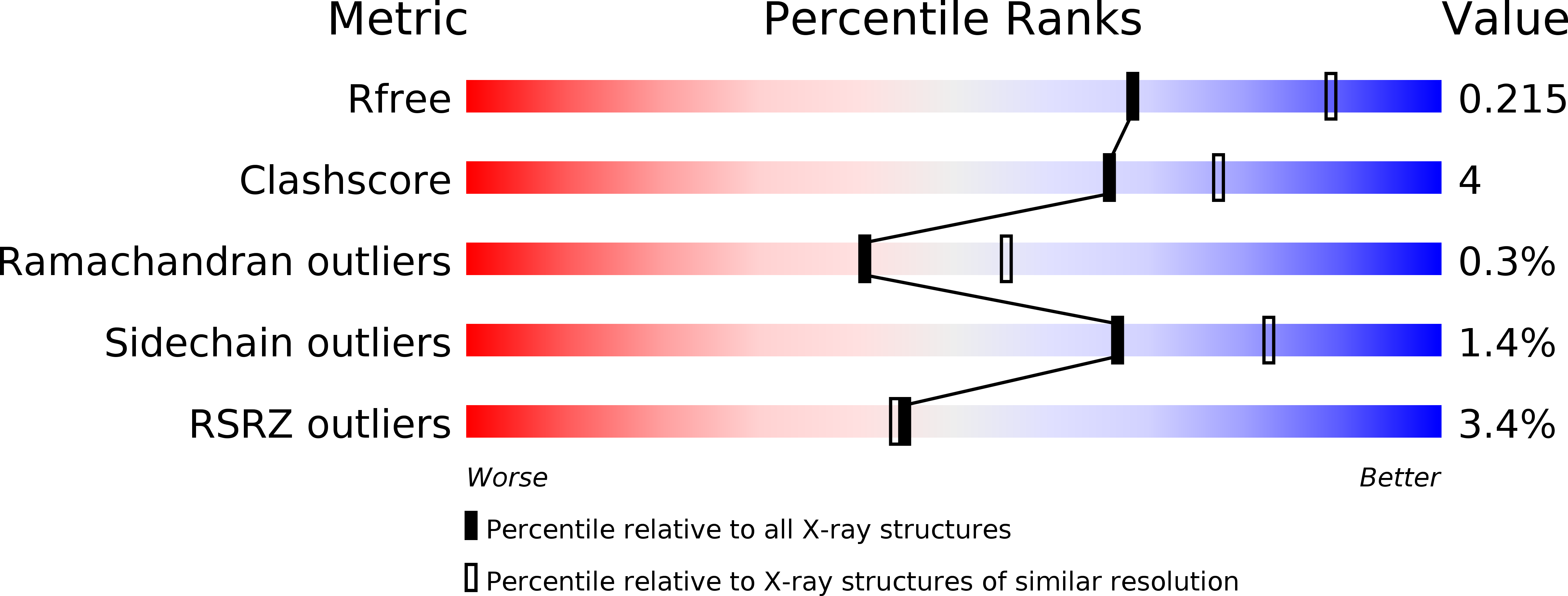
Resolution:
2.40 Å
R-Value Free:
0.21
R-Value Work:
0.17
R-Value Observed:
0.17
Space Group:
P 32 2 1
Macromolecular Entities
Structures with similar UniProt ID
Protein Blast
Polymer Type:polypeptide(L)
Molecule:Dihydroorotate dehydrogenase, mitochondrial
Gene (Uniprot):DHODH
Mutagens:N-terminus truncated
Chain IDs:A
Chain Length:367
Number of Molecules:1
Biological Source:Homo sapiens
Primary Citation
The structures of human dihydroorotate dehydrogenase with and without inhibitor reveal conformational flexibility in the inhibitor and substrate binding sites
Biochemistry 47 8929 8936 (2008)
PMID: 18672895 DOI: 10.1021/bi8003318

Abstact

Inhibitors of dihydroorotate dehydrogenase (DHODH) have been suggested for the treatment of rheumatoid arthritis, psoriasis, autoimmune diseases, Plasmodium, and bacterial and fungal infections. Here we present the structures of N-terminally truncated (residues Met30-Arg396) DHODH in complex with two inhibitors: a brequinar analogue (6) and a novel inhibitor (a fenamic acid derivative) (7), as well as the first structure of the enzyme to be characterized without any bound inhibitor. It is shown that 7 uses the "standard" brequinar binding mode and, in addition, interacts with Tyr356, a residue conserved in most class 2 DHODH proteins. Compared to the inhibitor-free structure, some of the amino acid side chains in the tunnel in which brequinar binds and which was suggested to be the binding site of ubiquinone undergo changes in conformation upon inhibitor binding. Using our data, the loop regions of residues Leu68-Arg72 and Asn212-Leu224, which were disordered in previously studied human DHODH structures, could be built into the electron density. The first of these loops, which is located at the entrance to the inhibitor-binding pocket, shows different conformations in the three structures, suggesting that it may interfere with inhibitor/cofactor binding. The second loop has been suggested to control the access of dihydroorotate to the active site of the enzyme and may be an important player in the enzymatic reaction. These observations provide new insights into the dynamic features of the DHODH reaction and suggest new approaches to the design of inhibitors against DHODH.

Legend

Protein

Chemical

Disease

Primary Citation of related structures
Feedback Form
Name
Email
Institute
Feedback