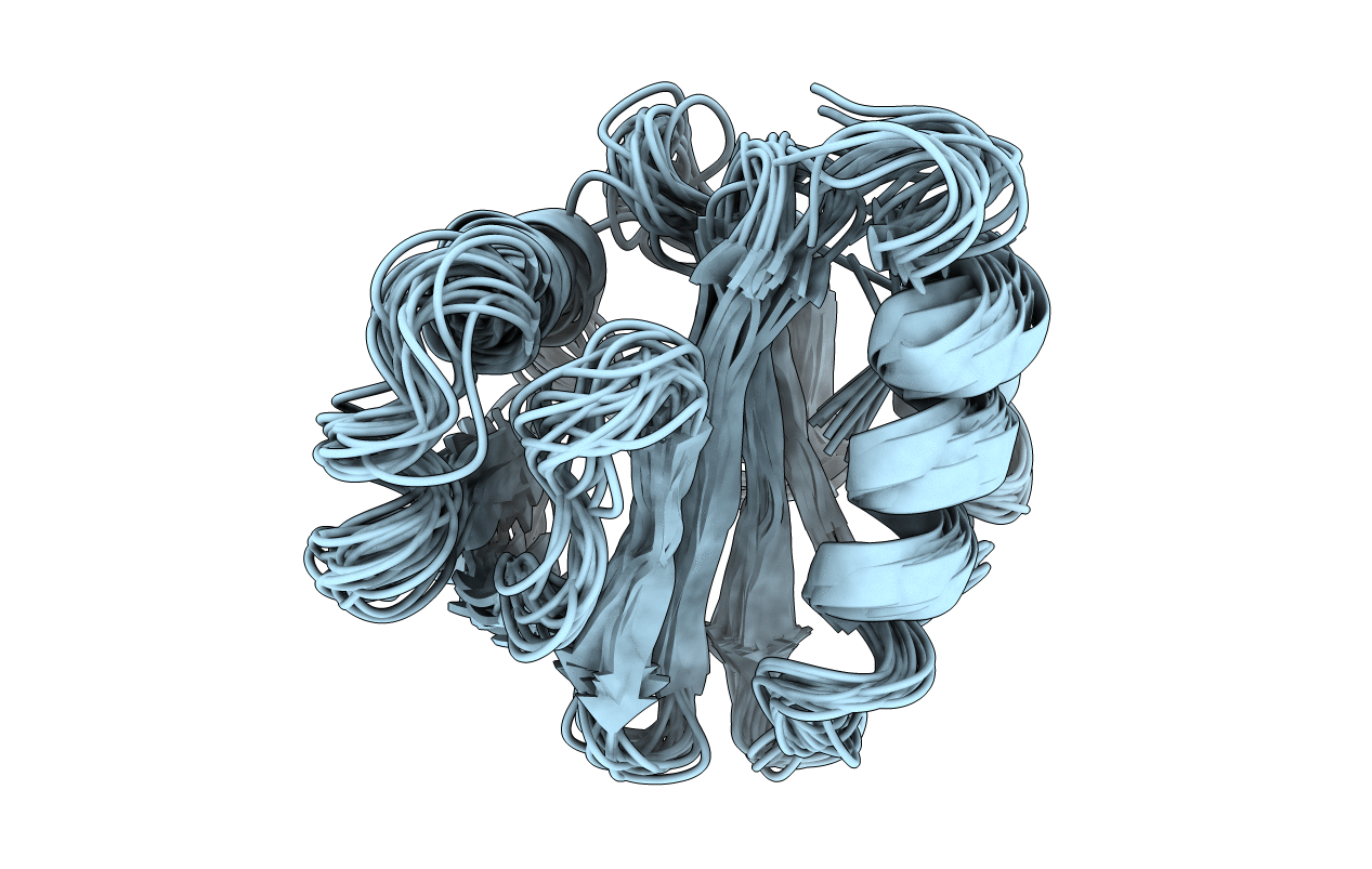
2PRF image
Deposition Date 1994-01-12
Release Date 1994-05-31
Last Version Date 2024-05-01
Entry Detail
PDB ID:
2PRF
Keywords:
Title:
THREE DIMENSIONAL SOLUTION STRUCTURE OF ACANTHAMOEBA PROFILIN I
Biological Source:
Source Organism:
Method Details:
Experimental Method:
Conformers Submitted:
19
Macromolecular Entities
Polymer Type:polypeptide(L)
Molecule:PROFILIN IA
Chain IDs:A
Chain Length:125
Number of Molecules:1
Biological Source:Acanthamoeba sp.
Ligand Molecules
Primary Citation
Three-dimensional solution structure of Acanthamoeba profilin-I.
J.Cell Biol. 122 1277 1283 (1993)
PMID: 8397216 DOI: 10.1083/jcb.122.6.1277

Abstact

We have determined a medium resolution three-dimensional solution structure of Acanthamoeba profilin-I by multidimensional nuclear magnetic resonance spectroscopy. This 13-kD actin binding protein consists of a five stranded antiparallel beta sheet flanked by NH2- and COOH-terminal helices on one face and by a third helix and a two stranded beta sheet on the other face. Data from actin-profilin cross-linking experiments and the localization of conserved residues between profilins in different phyla indicate that actin binding occurs on the molecular face occupied by the terminal helices. The other face of the molecule contains the residues that differ between Acanthamoeba profilins-I and II and may be important in determining the difference in polyphosphoinositide binding between these isoforms. This suggests that lipids and actin bind to different faces of the molecule.

Legend

Protein

Chemical

Disease

Primary Citation of related structures