2PRE image
Deposition Date 2007-05-04
Release Date 2008-01-22
Last Version Date 2024-10-16
Entry Detail
PDB ID:
2PRE
Keywords:
Title:
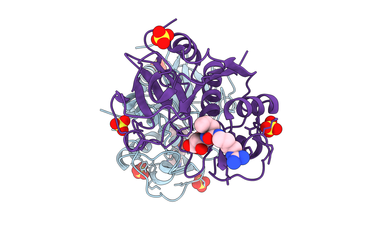
Crystal structure of plant cysteine protease Ervatamin-C complexed with irreversible inhibitor E-64 at 2.7 A resolution
Biological Source:
Source Organism:
Method Details:
Experimental Method:
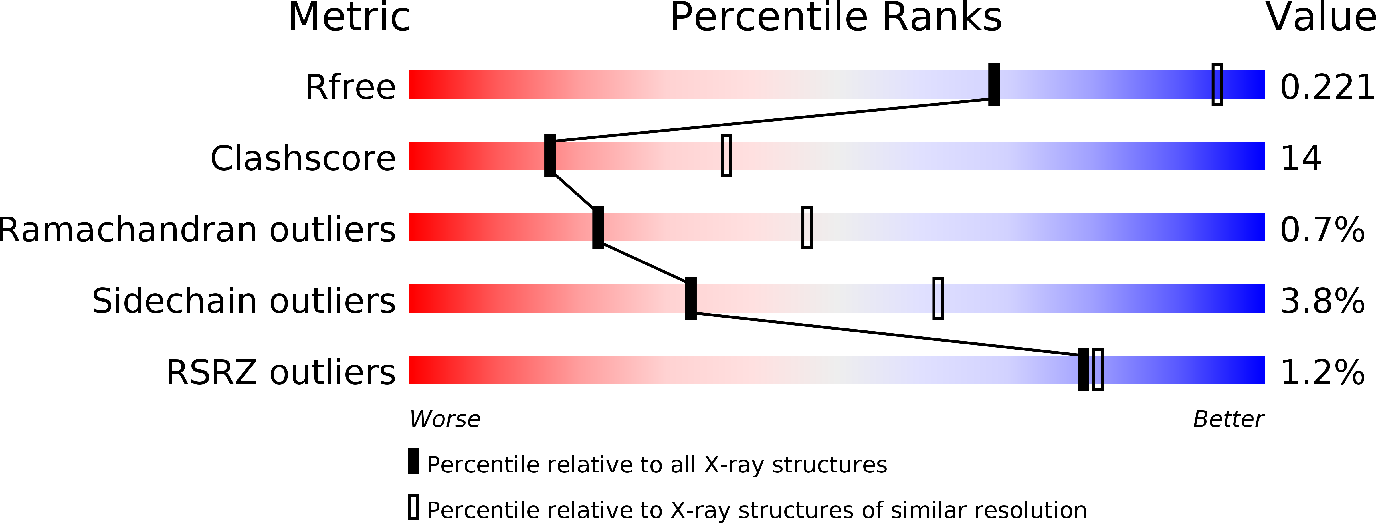
Resolution:
2.70 Å
R-Value Free:
0.23
R-Value Work:
0.19
R-Value Observed:
0.19
Space Group:
P 21 21 21
Macromolecular Entities
Polymer Type:polypeptide(L)
Molecule:Ervatamin-C
Chain IDs:A, B
Chain Length:208
Number of Molecules:2
Biological Source:Tabernaemontana divaricata
Primary Citation
Structural insights into the substrate specificity and activity of ervatamins, the papain-like cysteine proteases from a tropical plant, Ervatamia coronaria.
Febs J. 275 421 434 (2008)
PMID: 18167146 DOI: 10.1111/j.1742-4658.2007.06211.x

Abstact

Multiple proteases of the same family are quite often present in the same species in biological systems. These multiple proteases, despite having high homology in their primary and tertiary structures, show deviations in properties such as stability, activity, and specificity. It is of interest, therefore, to compare the structures of these multiple proteases in a single species to identify the structural changes, if any, that may be responsible for such deviations. Ervatamin-A, ervatamin-B and ervatamin-C are three such papain-like cysteine proteases found in the latex of the tropical plant Ervatamia coronaria, and are known not only for their high stability over a wide range of temperature and pH, but also for variations in activity and specificity among themselves and among other members of the family. Here we report the crystal structures of ervatamin-A and ervatamin-C, complexed with an irreversible inhibitor 1-[l-N-(trans-epoxysuccinyl)leucyl]amino-4-guanidinobutane (E-64), together with enzyme kinetics and molecular dynamic simulation studies. A comparison of these results with the earlier structures helps in a correlation of the structural features with the corresponding functional properties. The specificity constants (k(cat)/K(m)) for the ervatamins indicate that all of these enzymes have specificity for a branched hydrophobic residue at the P2 position of the peptide substrates, with different degrees of efficiency. A single amino acid change, as compared to ervatamin-C, in the S2 pocket of ervatamin-A (Ala67-->Tyr) results in a 57-fold increase in its k(cat)/K(m) value for a substrate having a Val at the P2 position. Our studies indicate a higher enzymatic activity of ervatamin-A, which has been subsequently explained at the molecular level from the three-dimensional structure of the enzyme and in the context of its helix polarizibility and active site plasticity.

Legend

Protein

Chemical

Disease

Primary Citation of related structures