Deposition Date
2007-04-26
Release Date
2007-12-04
Last Version Date
2024-05-22
Entry Detail
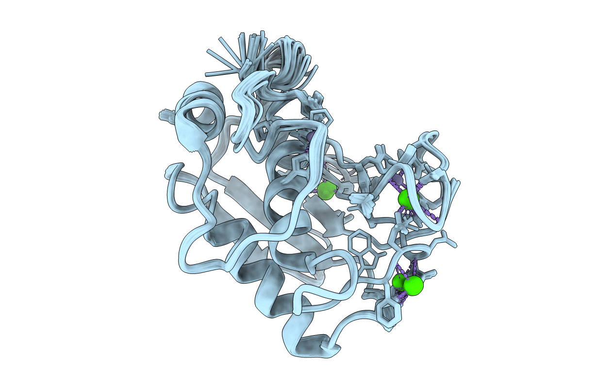
PDB ID:
2POJ
Keywords:
Title:
NMR Solution Structure of the Inhibitor-Free State of Macrophage Metalloelastase (MMP-12)
Biological Source:
Source Organism(s):
Homo sapiens (Taxon ID: 9606)
Expression System(s):
Method Details:
Experimental Method:
Conformers Calculated:
100
Conformers Submitted:
20
Selection Criteria:
structures with the lowest energy