
Deposition Date
2007-04-25
Release Date
2007-10-02
Last Version Date
2023-08-30
Entry Detail
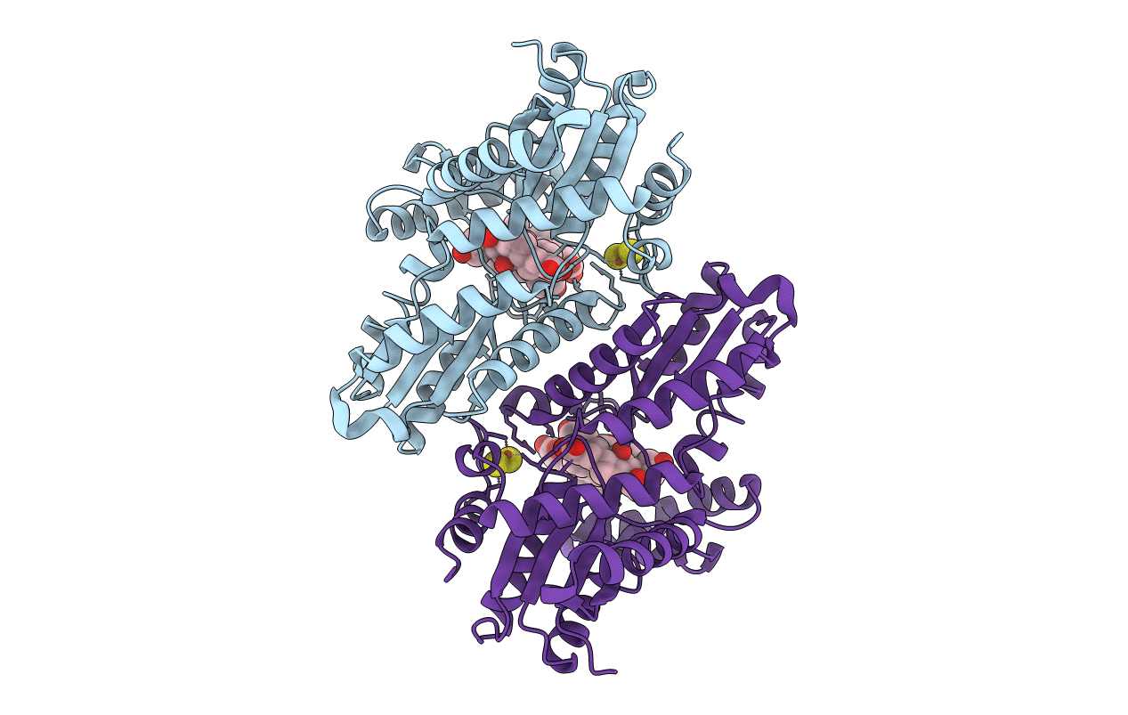
PDB ID:
2PO7
Keywords:
Title:
Crystal structure of human ferrochelatase mutant with His 341 replaced by Cys
Biological Source:
Source Organism(s):
Homo sapiens (Taxon ID: 9606)
Expression System(s):
Method Details:
Experimental Method:
Resolution:
2.20 Å
R-Value Free:
0.24
R-Value Work:
0.21
R-Value Observed:
0.21
Space Group:
P 21 21 21
