2PO4 image
Deposition Date 2007-04-25
Release Date 2008-04-01
Last Version Date 2024-02-21
Entry Detail
PDB ID:
2PO4
Keywords:
Title:
X-ray crystal structure of polymerase domain of the bacteriophage N4 virion RNA polymerase
Biological Source:
Source Organism:
Host Organism:
Method Details:
Experimental Method:
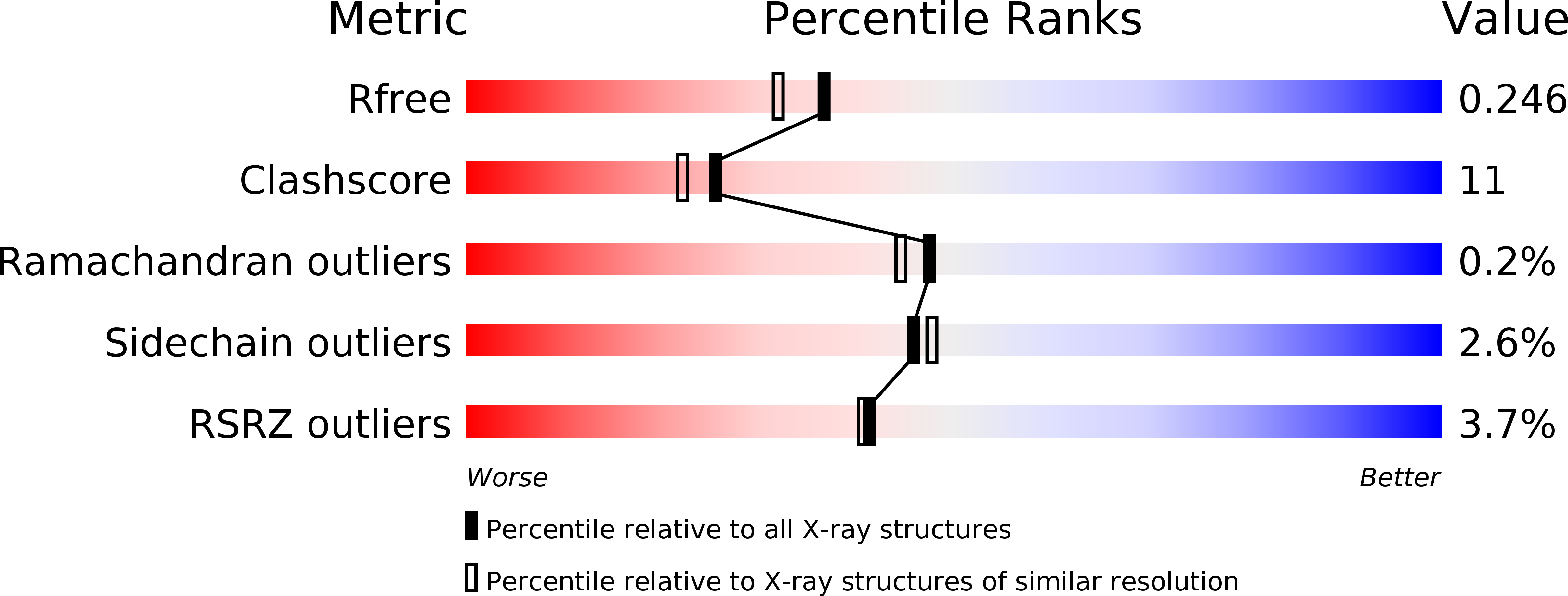
Resolution:
2.00 Å
R-Value Free:
0.24
R-Value Work:
0.21
Space Group:
P 21 21 21
Macromolecular Entities
Polymer Type:polypeptide(L)
Molecule:Virion RNA polymerase
Gene (Uniprot):50
Chain IDs:A
Chain Length:1104
Number of Molecules:1
Biological Source:Enterobacteria phage N4
Primary Citation
X-ray crystal structure of polymerase domain of the bacteriophage N4 virion RNA polymerase
Proc.Natl.Acad.Sci.USA 15 5046 5051 (2008)
PMID: 18362338 DOI: 10.1073/pnas.0712325105

Abstact

Coliphage N4 virion RNA polymerase (vRNAP), which is injected into the host upon infection, transcribes the phage early genes from promoters that have a 5-bp stem-3 nt loop hairpin structure. Here, we describe the 2.0-A resolution x-ray crystal structure of N4 mini-vRNAP, a member of the T7-like, single-unit RNAP family and the minimal component having all RNAP functions of the full-length vRNAP. The structure resembles a "fisted right hand" with Fingers, Palm and Thumb subdomains connected to an N-terminal domain. We established that the specificity loop extending from the Fingers along with W129 of the N-terminal domain play critical roles in hairpin-promoter recognition. A comparison with the structure of the T7 RNAP initiation complex reveals that the pathway of the DNA to the active site is blocked in the apo-form vRNAP, indicating that vRNAP must undergo a large-scale conformational change upon promoter DNA binding and explaining the highly restricted promoter specificity of vRNAP that is essential for phage early transcription.

Legend

Protein

Chemical

Disease

Primary Citation of related structures