2PNL image
Deposition Date 2007-04-24
Release Date 2007-06-05
Last Version Date 2024-10-30
Entry Detail
PDB ID:
2PNL
Keywords:
Title:
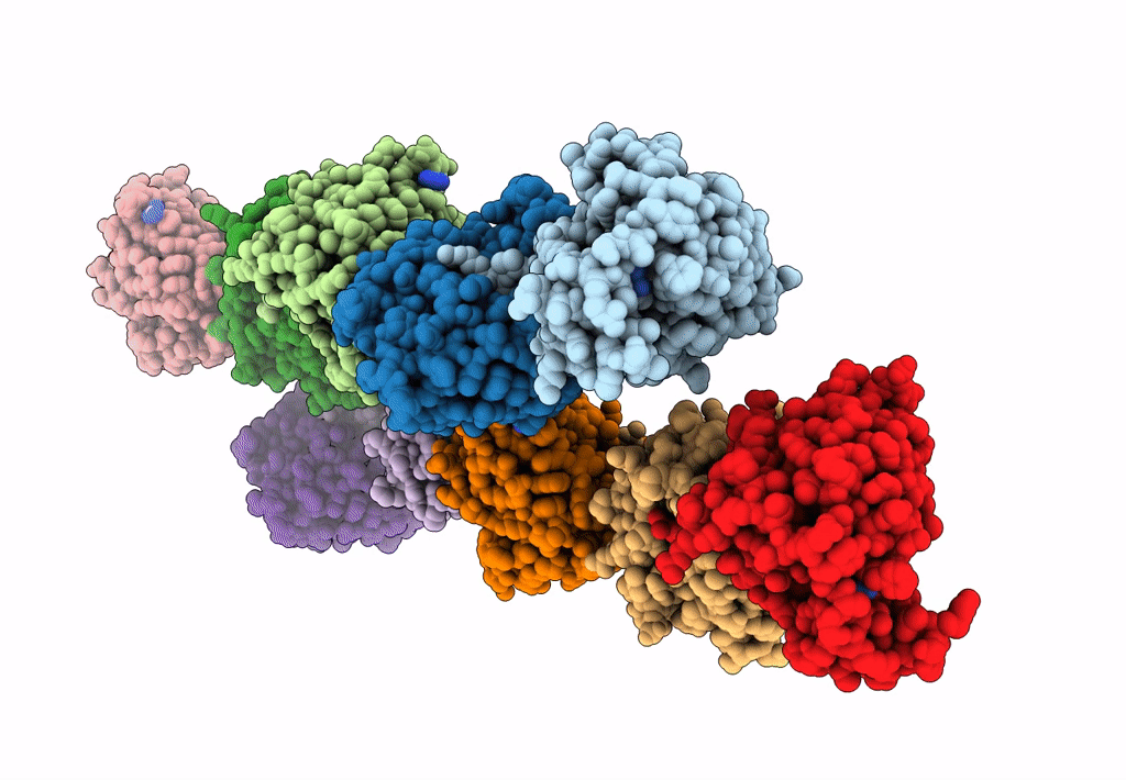
Crystal structure of VP4 protease from infectious pancreatic necrosis virus (IPNV) in space group P1
Biological Source:
Source Organism:
Method Details:
Experimental Method:
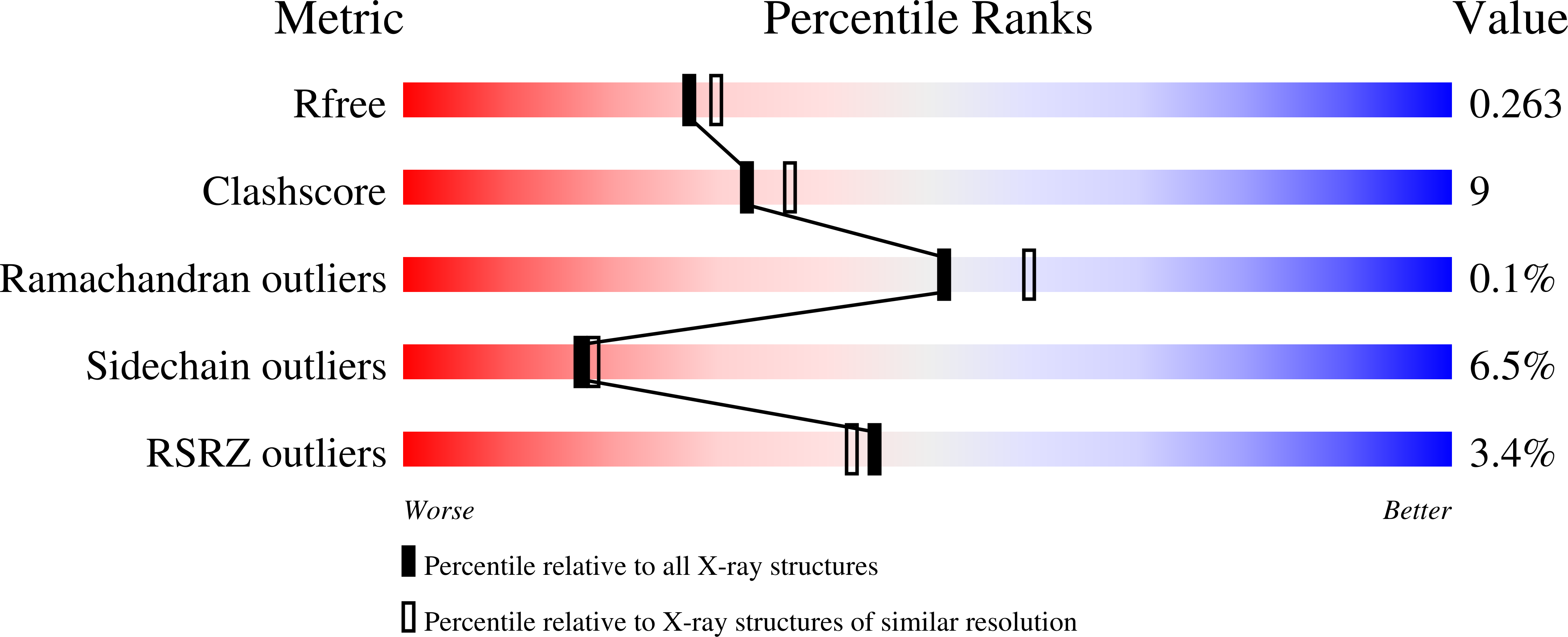
Resolution:
2.21 Å
R-Value Free:
0.26
R-Value Work:
0.19
R-Value Observed:
0.19
Space Group:
P 1
Macromolecular Entities
Polymer Type:polypeptide(L)
Molecule:Protease VP4
Mutations:K674A
Chain IDs:A, B, C, D, E, F, G, H, I, J
Chain Length:203
Number of Molecules:10
Biological Source:Infectious pancreatic necrosis virus
Modified Residue
Compound ID Chain ID Parent Comp ID Details 2D Image
MSE A MET SELENOMETHIONINE
Ligand Molecules
Primary Citation
Crystal structure of the VP4 protease from infectious pancreatic necrosis virus reveals the acyl-enzyme complex for an intermolecular self-cleavage reaction.
J.Biol.Chem. 282 24928 24937 (2007)
PMID: 17553791 DOI: 10.1074/jbc.M701551200

Abstact

Infectious pancreatic necrosis virus (IPNV), an aquatic birnavirus that infects salmonid fish, encodes a large polyprotein (NH(2)-pVP2-VP4-VP3-COOH) that is processed through the proteolytic activity of its own protease, VP4, to release the proteins pVP2 and VP3. pVP2 is further processed to give rise to the capsid protein VP2 and three peptides that are incorporated into the virion. Reported here are two crystal structures of the IPNV VP4 protease solved from two different crystal symmetries. The electron density at the active site in the triclinic crystal form, refined to 2.2-A resolution, reveals the acyl-enzyme complex formed with an internal VP4 cleavage site. The complex was generated using a truncated enzyme in which the general base lysine was substituted. Inside the complex, the nucleophilic Ser(633)Ogamma forms an ester bond with the main-chain carbonyl of the C-terminal residue, Ala(716), of a neighboring VP4. The structure of this substrate-VP4 complex allows us to identify the S1, S3, S5, and S6 substrate binding pockets as well as other substrate-VP4 interactions and therefore provides structural insights into the substrate specificity of this enzyme. The structure from the hexagonal crystal form, refined to 2.3-A resolution, reveals the free-binding site of the protease. Three-dimensional alignment with the VP4 of blotched snakehead virus, another birnavirus, shows that the overall structure of VP4 is conserved despite a low level of sequence identity (approximately 19%). The structure determinations of IPNV VP4, the first of an acyl-enzyme complex for a Ser/Lys dyad protease, provide insights into the catalytic mechanism and substrate recognition of this type of protease.

Legend

Protein

Chemical

Disease

Primary Citation of related structures