2PMC image
Entry Detail
PDB ID:
2PMC
Title:
Crystal Structure of CheY-Mg(2+) in Complex with CheZ(C15) Peptide solved from a P1 Crystal
Biological Source:
Source Organism:
Host Organism:
PDB Version:
Deposition Date:
2007-04-20
Release Date:
2008-01-15
Method Details:
Experimental Method:
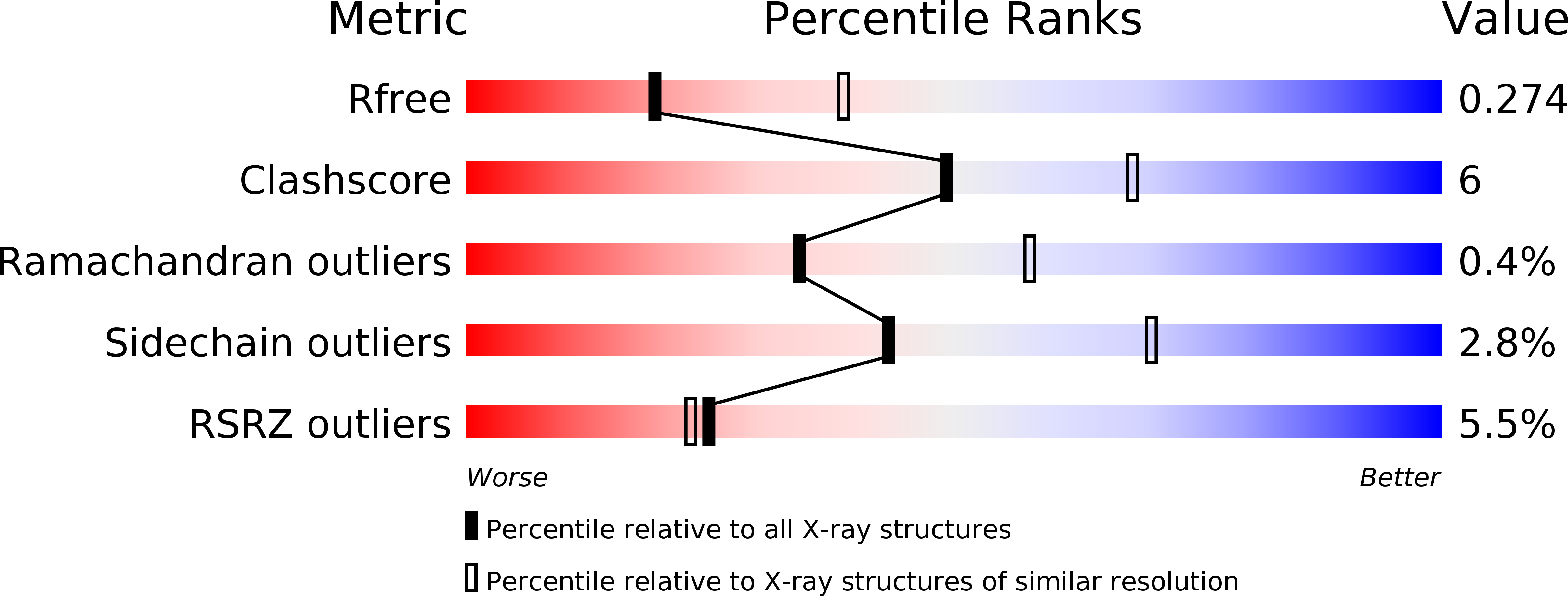
Resolution:
2.69 Å
R-Value Free:
0.28
R-Value Work:
0.20
R-Value Observed:
0.21
Space Group:
P 1
Macromolecular Entities
Polymer Type:polypeptide(L)
Description:Chemotaxis protein cheY
Chain IDs:A, B, C, D
Chain Length:128
Number of Molecules:4
Biological Source:Salmonella typhimurium
Polymer Type:polypeptide(L)
Description:Chemotaxis protein cheZ
Chain IDs:E, F
Chain Length:15
Number of Molecules:2
Biological Source:
Ligand Molecules
Primary Citation
Interaction of CheY with the C-terminal peptide of CheZ.
J.Bacteriol. 190 1419 1428 (2008)
PMID: 18083806 DOI: 10.1128/JB.01414-07

Abstact

Chemotaxis, a means for motile bacteria to sense the environment and achieve directed swimming, is controlled by flagellar rotation. The primary output of the chemotaxis machinery is the phosphorylated form of the response regulator CheY (P-CheY). The steady-state level of P-CheY dictates the direction of rotation of the flagellar motor. The chemotaxis signal in the form of P-CheY is terminated by the phosphatase CheZ. Efficient dephosphorylation of CheY by CheZ requires two distinct protein-protein interfaces: one involving the strongly conserved C-terminal helix of CheZ (CheZ(C)) tethering the two proteins together and the other constituting an active site for catalytic dephosphorylation. In a previous work (J. Guhaniyogi, V. L. Robinson, and A. M. Stock, J. Mol. Biol. 359:624-645, 2006), we presented high-resolution crystal structures of CheY in complex with the CheZ(C) peptide that revealed alternate binding modes subject to the conformational state of CheY. In this study, we report biochemical and structural data that support the alternate-binding-mode hypothesis and identify key recognition elements in the CheY-CheZ(C) interaction. In addition, we present kinetic studies of the CheZ(C)-associated effect on CheY phosphorylation with its physiologically relevant phosphodonor, the histidine kinase CheA. Our results indicate mechanistic differences in phosphotransfer from the kinase CheA versus that from small-molecule phosphodonors, explaining a modest twofold increase of CheY phosphorylation with the former, observed in this study, relative to a 10-fold increase previously documented with the latter.

Legend

Protein

Chemical

Disease

Primary Citation of related structures