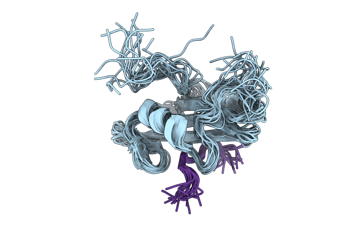
2PLE image
Entry Detail
PDB ID:
2PLE
Title:
NUCLEAR MAGNETIC RESONANCE STRUCTURE OF AN SH2 DOMAIN OF PHOSPHOLIPASE C-GAMMA1 COMPLEXED WITH A HIGH AFFINITY BINDING PEPTIDE
Biological Source:
Source Organism:
PDB Version:
Deposition Date:
1994-08-19
Release Date:
1995-01-26
Method Details:
Experimental Method:
Conformers Submitted:
18
Macromolecular Entities
Polymer Type:polypeptide(L)
Description:PHOSPHOLIPASE C GAMMA-1, C-TERMINAL SH2 DOMAIN
Chain IDs:A
Chain Length:105
Number of Molecules:1
Biological Source:Bos taurus
Polymer Type:polypeptide(L)
Description:PHOSPHOPEPTIDE FROM PDGF
Chain IDs:B
Chain Length:12
Number of Molecules:1
Biological Source:Bos taurus
Modified Residue
Compound ID Chain ID Parent Comp ID Details 2D Image
PTR B TYR O-PHOSPHOTYROSINE
Ligand Molecules
Primary Citation
Nuclear magnetic resonance structure of an SH2 domain of phospholipase C-gamma 1 complexed with a high affinity binding peptide.
Cell(Cambridge,Mass.) 77 461 472 (1994)
PMID: 8181064 DOI: 10.1016/0092-8674(94)90160-0

Abstact

The solution structure of the C-terminal SH2 domain of phospholipase C-gamma 1 (PLC-gamma 1), in complex with a phosphopeptide corresponding to its Tyr-1021 high affinity binding site on the platelet-derived growth factor receptor, has been determined by nuclear magnetic resonance spectroscopy. The topology of the SH2-phosphopeptide complex is similar to previously reported Src and Lck SH2 complexes. However, the binding site for residues C-terminal to the phosphotyrosine (pTyr) is an extended groove that contacts peptide residues at the +1 to +6 positions relative to the pTyr. This striking difference from Src and Lck reflects the fact that the PLC-gamma 1 complex involves binding of a phosphopeptide with predominantly hydrophobic residues C-terminal to the pTyr and therefore serves as a prototype for a second class of SH2-phosphopeptide interactions.

Legend

Protein

Chemical

Disease

Primary Citation of related structures