2PL7 image
Deposition Date 2007-04-19
Release Date 2007-07-17
Last Version Date 2024-11-13
Entry Detail
PDB ID:
2PL7
Title:
Orhorhombic crystal structure of hydrophobin HFBII in the presence of a detergent
Biological Source:
Source Organism:
Method Details:
Experimental Method:
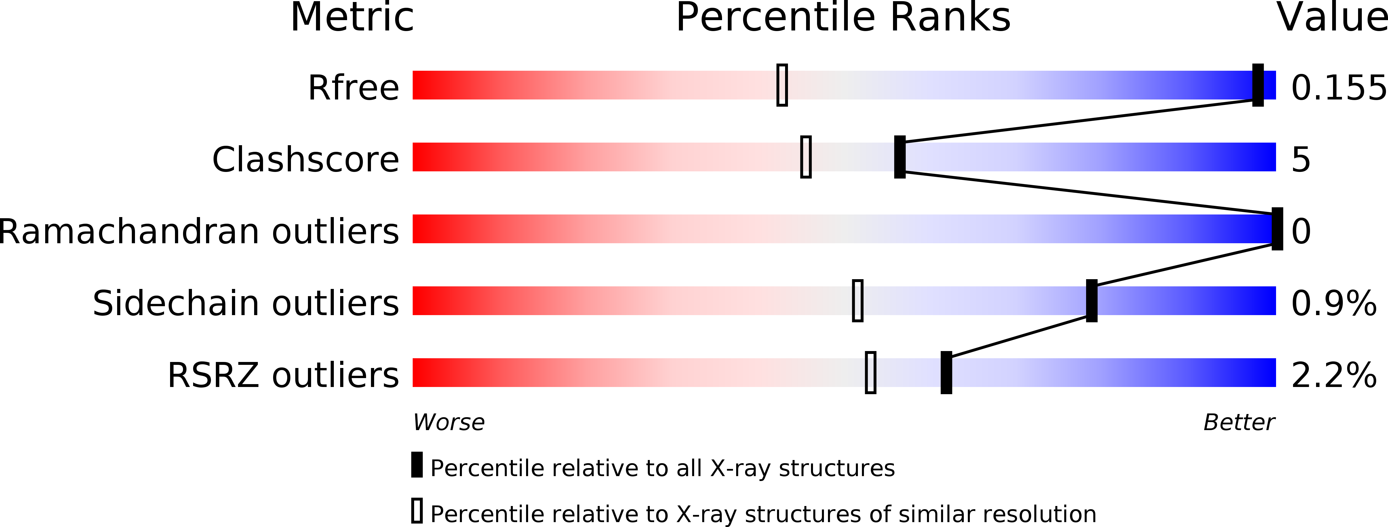
Resolution:
1.00 Å
R-Value Free:
0.15
R-Value Observed:
0.17
Space Group:
P 21 21 21
Macromolecular Entities
Polymer Type:polypeptide(L)
Molecule:Hydrophobin-2
Gene (Uniprot):hfb2
Chain IDs:A, B
Chain Length:71
Number of Molecules:2
Biological Source:Hypocrea jecorina
Primary Citation
Crystal Structures of Hydrophobin HFBII in the Presence of Detergent Implicate the Formation of Fibrils and Monolayer Films.
J.Biol.Chem. 282 28733 28739 (2007)
PMID: 17636262 DOI: 10.1074/jbc.M704238200

Abstact

Hydrophobins are small, amphiphilic proteins secreted by filamentous fungi. Their functionality arises from a patch of hydrophobic residues on the protein surface. Spontaneous self-assembly of hydrophobins leads to the formation of an amphiphilic layer that remarkably reduces the surface tension of water. We have determined by x-ray diffraction two new crystal structures of Trichoderma reesei hydrophobin HFBII in the presence of a detergent. The monoclinic crystal structure (2.2A resolution, R = 22, R(free) = 28) is composed of layers of hydrophobin molecules where the hydrophobic surface areas of the molecules are aligned within the layer. Viewed perpendicular to the aligned hydrophobic surface areas, the molecules in the layer pack together to form six-membered rings, thus leaving small pores in the layer. Similar packing has been observed in the atomic force microscopy images of the self-assembled layers of class II hydrophobin, indicating that the crystal structure resembles that of natural hydrophobin film. The orthorhombic crystal structure (1.0 A resolution, R = 13, R(free) = 15) is composed of fiber-like arrays of protein molecules. Rodlet structures have been observed on amphiphilic layers formed by class I hydrophobins; fibrils of class II hydrophobins appear by vigorous shaking. We propose that the structure of the fibrils and/or rodlets is similar to that observed in the crystal structure.

Legend

Protein

Chemical

Disease

Primary Citation of related structures