Deposition Date
1993-06-04
Release Date
1994-01-31
Last Version Date
2024-10-16
Entry Detail
PDB ID:
2PKC
Keywords:
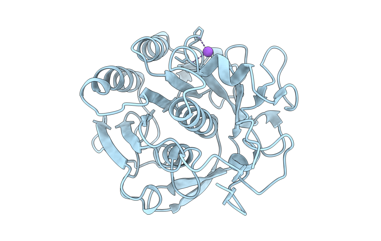
Title:
CRYSTAL STRUCTURE OF CALCIUM-FREE PROTEINASE K AT 1.5 ANGSTROMS RESOLUTION
Biological Source:
Source Organism(s):
Engyodontium album (Taxon ID: 37998)
Method Details:
Experimental Method:
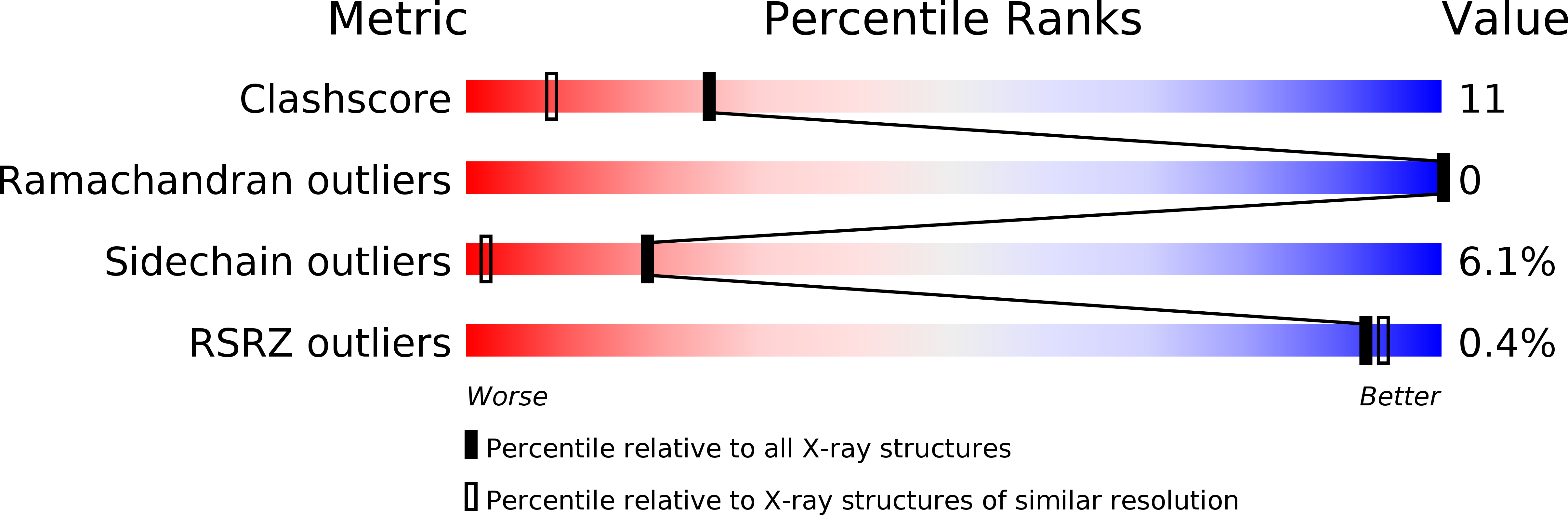
Resolution:
1.50 Å
R-Value Observed:
0.20
Space Group:
P 43 21 2