
Deposition Date
2007-04-17
Release Date
2008-03-25
Last Version Date
2023-08-30
Entry Detail
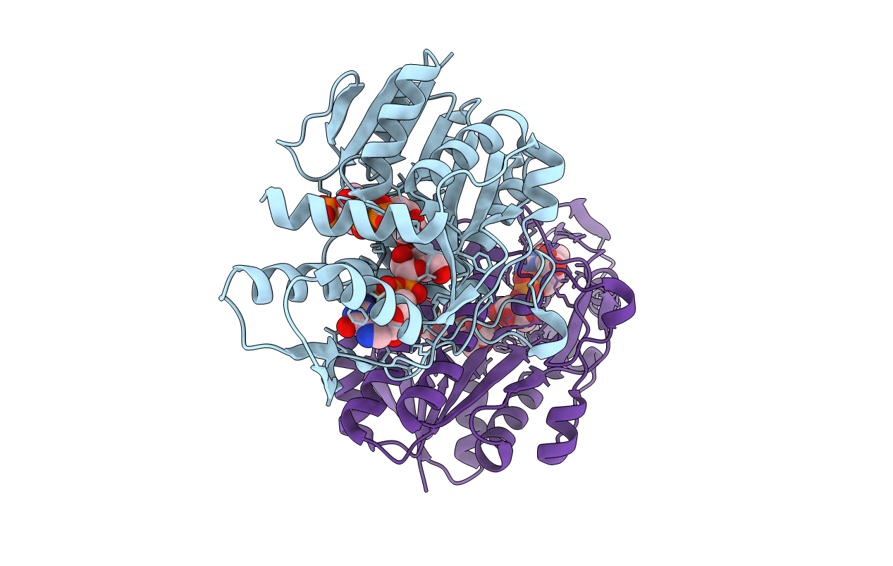
PDB ID:
2PK3
Keywords:
Title:
Crystal Structure of a GDP-4-keto-6-deoxy-D-mannose reductase
Biological Source:
Source Organism(s):
Aneurinibacillus thermoaerophilus (Taxon ID: 143495)
Expression System(s):
Method Details:
Experimental Method:
Resolution:
1.82 Å
R-Value Free:
0.19
R-Value Work:
0.16
R-Value Observed:
0.16
Space Group:
P 1
