2PK0 image
Deposition Date 2007-04-17
Release Date 2007-06-12
Last Version Date 2024-04-03
Entry Detail
PDB ID:
2PK0
Title:
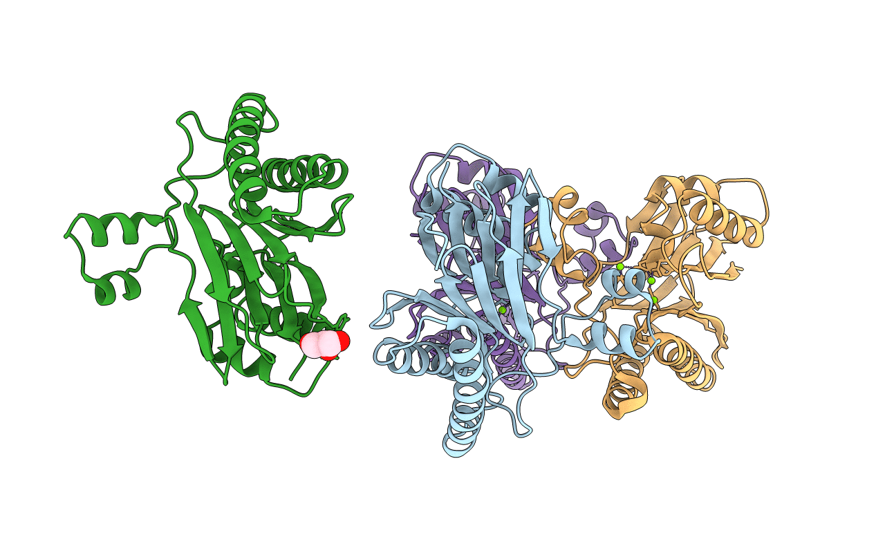
Structure of the S. agalactiae serine/threonine phosphatase at 2.65 resolution
Biological Source:
Source Organism:
Method Details:
Experimental Method:
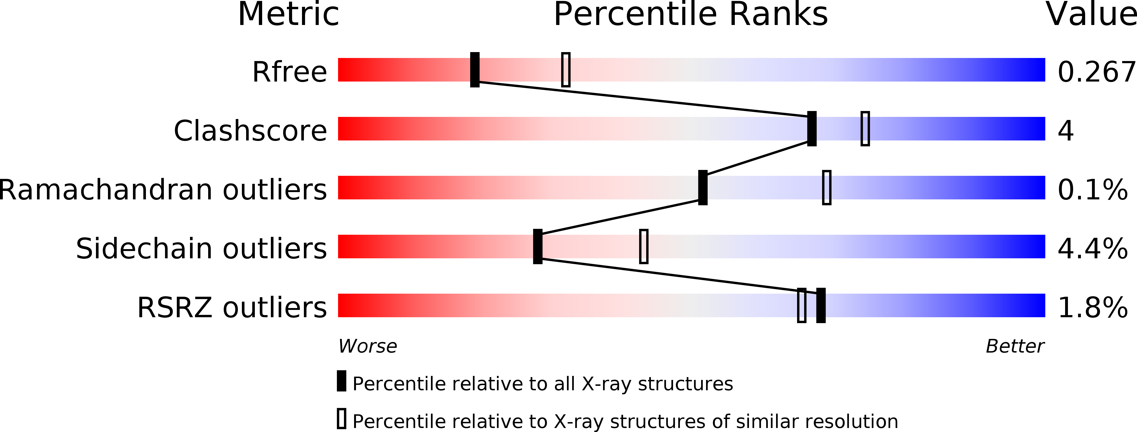
Resolution:
2.65 Å
R-Value Free:
0.27
R-Value Work:
0.19
R-Value Observed:
0.20
Space Group:
P 21 21 2
Macromolecular Entities
Structures with similar UniProt ID
Protein Blast
Polymer Type:polypeptide(L)
Molecule:Serine/threonine protein phosphatase Stp1
Chain IDs:A, B, C, D
Chain Length:250
Number of Molecules:4
Biological Source:Streptococcus agalactiae
Primary Citation
Structure of Streptococcus agalactiae serine/threonine phosphatase. The subdomain conformation is coupled to the binding of a third metal ion
Febs J. 274 3128 3137 (2007)
PMID: 17521332 DOI: 10.1111/j.1742-4658.2007.05845.x

Abstact

We solved the crystal structure of Streptococcus agalactiae serine/threonine phosphatase (SaSTP) using a combination of single-wavelength anomalous dispersion phasing and molecular replacement. The overall structure resembles that of previously characterized members of the PPM/PP2C STP family. The asymmetric unit contains four monomers and we observed two novel conformations for the flap domain among them. In one of these conformations, the enzyme binds three metal ions, whereas in the other it binds only two. The three-metal ion structure also has the active site arginine in a novel conformation. The switch between the two- and three-metal ion structures appears to be binding of another monomer to the active site of STP, which promotes binding of the third metal ion. This interaction may mimic the binding of a product complex, especially since the motif binding to the active site contains a serine residue aligning remarkably well with the phosphate found in the human STP structure.

Legend

Protein

Chemical

Disease

Primary Citation of related structures