2PID image
Deposition Date 2007-04-13
Release Date 2007-10-23
Last Version Date 2024-03-13
Entry Detail
PDB ID:
2PID
Keywords:
Title:
Crystal structure of human mitochondrial tyrosyl-tRNA synthetase in complex with an adenylate analog
Biological Source:
Source Organism:
Homo sapiens (Taxon ID: 9606)
Host Organism:
Method Details:
Experimental Method:
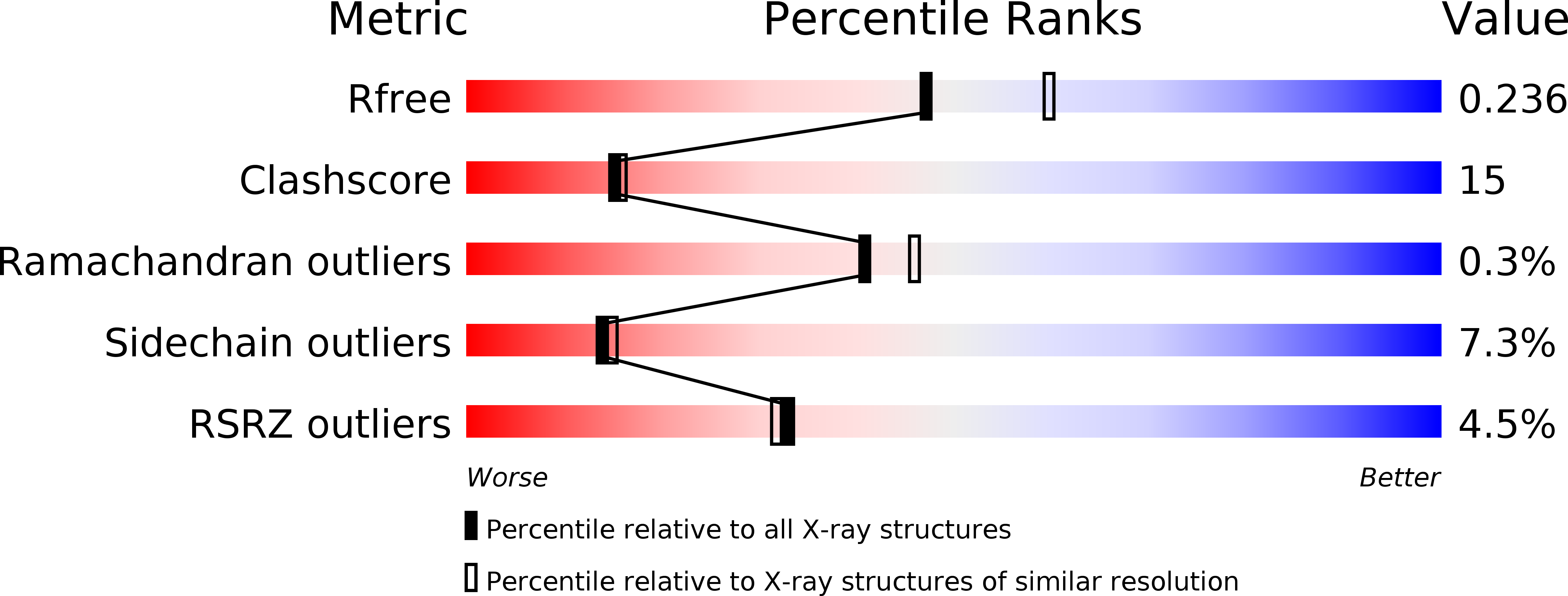
Resolution:
2.20 Å
R-Value Free:
0.24
R-Value Work:
0.19
R-Value Observed:
0.19
Space Group:
P 21 21 21
Macromolecular Entities
Polymer Type:polypeptide(L)
Molecule:Tyrosyl-tRNA synthetase
Gene (Uniprot):YARS2
Chain IDs:A, B
Chain Length:356
Number of Molecules:2
Biological Source:Homo sapiens
Ligand Molecules
Primary Citation
Crystal Structure of Human Mitochondrial Tyrosyl-tRNA Synthetase Reveals Common and Idiosyncratic Features.
Structure 15 1505 1516 (2007)
PMID: 17997975 DOI: 10.1016/j.str.2007.09.018

Abstact

We report the structure of a strictly mitochondrial human synthetase, namely tyrosyl-tRNA synthetase (mt-TyrRS), in complex with an adenylate analog at 2.2 A resolution. The structure is that of an active enzyme deprived of the C-terminal S4-like domain and resembles eubacterial TyrRSs with a canonical tyrosine-binding pocket and adenylate-binding residues typical of class I synthetases. Two bulges at the enzyme surface, not seen in eubacterial TyrRSs, correspond to conserved sequences in mt-TyrRSs. The synthetase electrostatic surface potential differs from that of other TyrRSs, including the human cytoplasmic homolog and the mitochondrial one from Neurospora crassa. The homodimeric human mt-TyrRS shows an asymmetry propagating from the dimer interface toward the two catalytic sites and extremities of each subunit. Mutagenesis of the catalytic domain reveals functional importance of Ser200 in line with an involvement of A73 rather than N1-N72 in tyrosine identity.

Legend

Protein

Chemical

Disease

Primary Citation of related structures