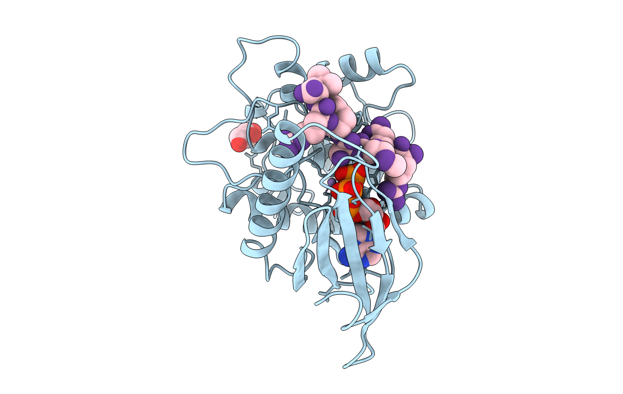
2PHK image
Deposition Date 1998-06-18
Release Date 1999-01-13
Last Version Date 2024-05-22
Entry Detail
PDB ID:
2PHK
Title:
THE CRYSTAL STRUCTURE OF A PHOSPHORYLASE KINASE PEPTIDE SUBSTRATE COMPLEX: KINASE SUBSTRATE RECOGNITION
Biological Source:
Source Organism:
Method Details:
Experimental Method:
Resolution:
2.60 Å
R-Value Free:
0.30
R-Value Work:
0.23
R-Value Observed:
0.25
Space Group:
P 32 2 1
Macromolecular Entities
Structures with similar UniProt ID
Protein Blast
Polymer Type:polypeptide(L)
Molecule:PHOSPHORYLASE KINASE
Gene (Uniprot):PHKG1
Chain IDs:A
Chain Length:277
Number of Molecules:1
Biological Source:Oryctolagus cuniculus
Primary Citation
The crystal structure of a phosphorylase kinase peptide substrate complex: kinase substrate recognition.
EMBO J. 16 6646 6658 (1997)
PMID: 9362479 DOI: 10.1093/emboj/16.22.6646

Abstact

The structure of a truncated form of the gamma-subunit of phosphorylase kinase (PHKgammat) has been solved in a ternary complex with a non-hydrolysable ATP analogue (adenylyl imidodiphosphate, AMPPNP) and a heptapeptide substrate related in sequence to both the natural substrate and to the optimal peptide substrate. Kinetic characterization of the phosphotransfer reaction confirms the peptide to be a good substrate, and the structure allows identification of key features responsible for its high affinity. Unexpectedly, the substrate peptide forms a short anti-parallel beta-sheet with the kinase activation segment, the region which in other kinases plays an important role in regulation of enzyme activity. This anchoring of the main chain of the substrate peptide at a fixed distance from the gamma-phosphate of ATP explains the selectivity of PHK for serine/threonine over tyrosine as a substrate. The catalytic core of PHK exists as a dimer in crystals of the ternary complex, and the relevance of this phenomenon to its in vivo recognition of dimeric glycogen phosphorylase b is considered.

Legend

Protein

Chemical

Disease

Primary Citation of related structures