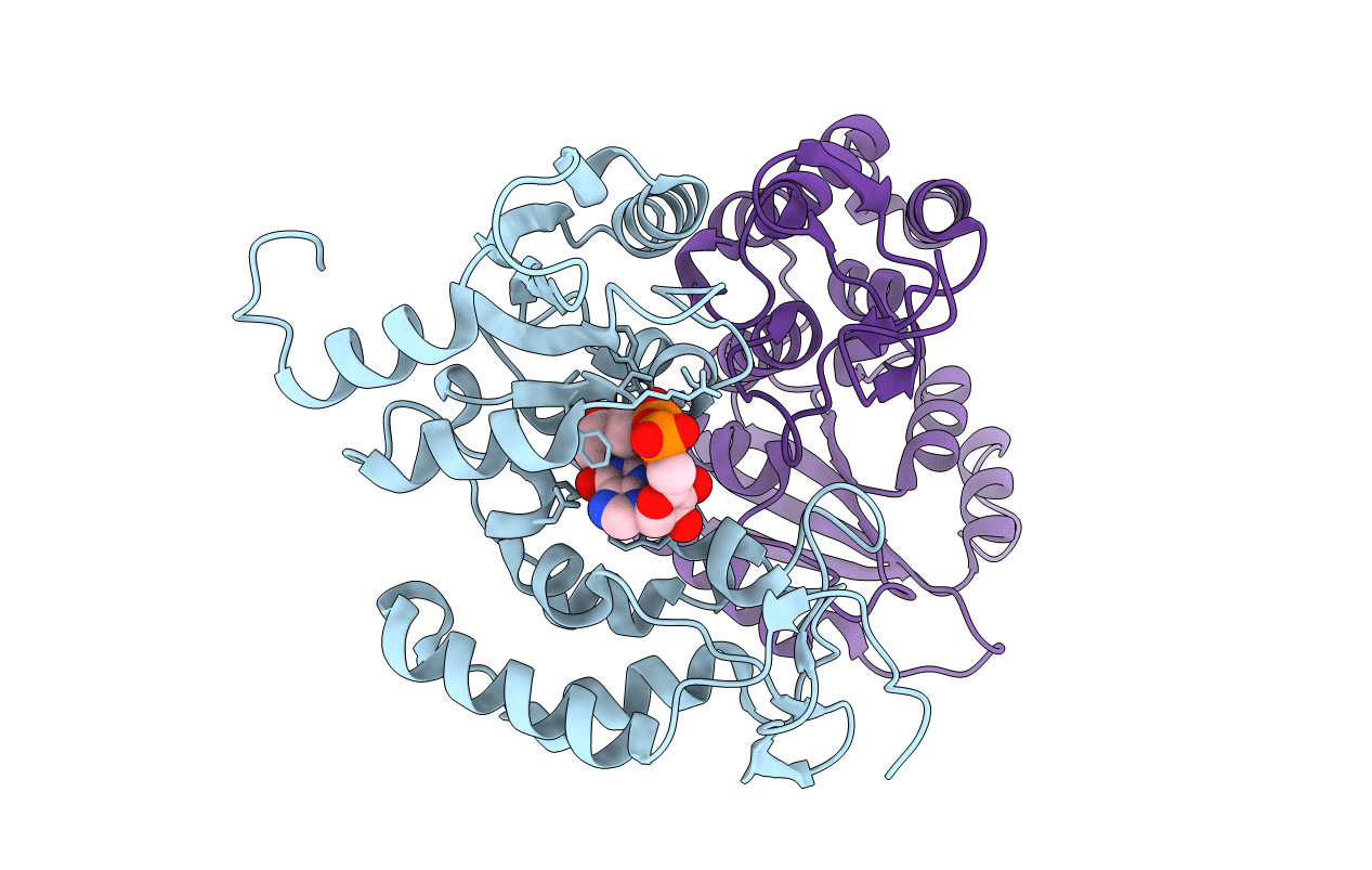
2PGJ image
Deposition Date 2007-04-09
Release Date 2007-04-24
Last Version Date 2024-11-20
Entry Detail
PDB ID:
2PGJ
Keywords:
Title:
Catalysis associated conformational changes revealed by human cd38 complexed with a non-hydrolyzable substrate analog
Biological Source:
Source Organism:
Homo sapiens (Taxon ID: 9606)
Host Organism:
Method Details:
Experimental Method:
Resolution:
1.71 Å
R-Value Free:
0.22
R-Value Work:
0.19
R-Value Observed:
0.19
Space Group:
P 1
Macromolecular Entities
Polymer Type:polypeptide(L)
Molecule:ADP-ribosyl cyclase 1
Gene (Uniprot):CD38
Mutations:Q49T, N100D,N164D, N209D, N219D
Chain IDs:A, B
Chain Length:262
Number of Molecules:2
Biological Source:Homo sapiens
Ligand Molecules
Primary Citation
Catalysis-associated Conformational Changes Revealed by Human CD38 Complexed with a Non-hydrolyzable Substrate Analog
J.Biol.Chem. 282 24825 24832 (2007)
PMID: 17591784 DOI: 10.1074/jbc.M701653200

Abstact

Cyclic ADP-ribose (cADPR) is a calcium mobilization messenger important for mediating a wide range of physiological functions. The endogenous levels of cADPR in mammalian tissues are primarily controlled by CD38, a multifunctional enzyme capable of both synthesizing and hydrolyzing cADPR. In this study, a novel non-hydrolyzable analog of cADPR, N1-cIDPR (N1-cyclic inosine diphosphate ribose), was utilized to elucidate the structural determinants involved in the hydrolysis of cADPR. N1-cIDPR inhibits CD38-catalyzed cADPR hydrolysis with an IC(50) of 0.26 mM. N1-cIDPR forms a complex with CD38 or its inactive mutant in which the catalytic residue Glu-226 is mutated. Both complexes have been determined by x-ray crystallography at 1.7 and 1.76 A resolution, respectively. The results show that N1-cIDPR forms two hydrogen bonds (2.61 and 2.64 A) with Glu-226, confirming our previously proposed model for cADPR catalysis. Structural analyses reveal that both the enzyme and substrate cADPR undergo catalysis-associated conformational changes. From the enzyme side, residues Glu-146, Asp-147, and Trp-125 work collaboratively to facilitate the formation of the Michaelis complex. From the substrate side, cADPR is found to change its conformation to fit into the active site until it reaches the catalytic residue. The binary CD38-cADPR model described here represents the most detailed description of the CD38-catalyzed hydrolysis of cADPR at atomic resolution. Our structural model should provide insights into the design of effective cADPR analogs.

Legend

Protein

Chemical

Disease

Primary Citation of related structures