Deposition Date
1988-01-25
Release Date
1989-01-09
Last Version Date
2024-02-21
Entry Detail
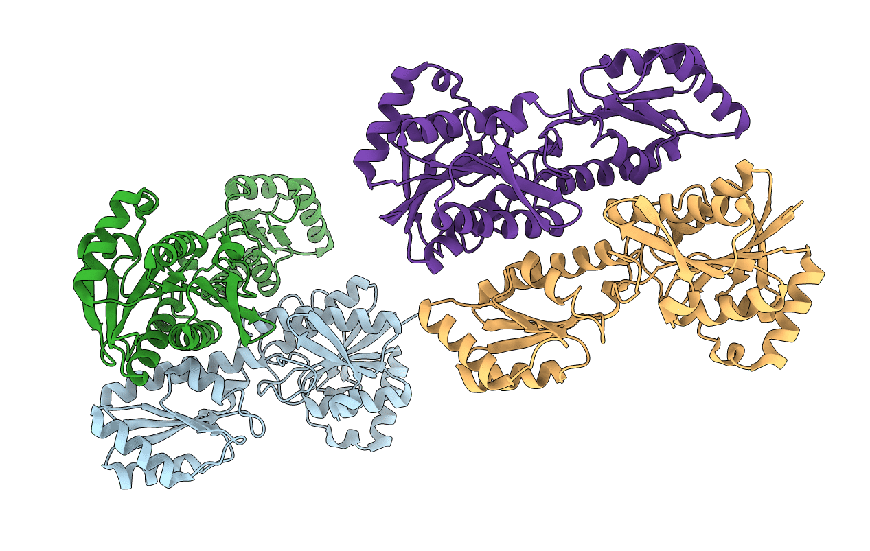
PDB ID:
2PFK
Keywords:
Title:
THE CRYSTAL STRUCTURE OF UNLIGANDED PHOSPHOFRUCTOKINASE FROM ESCHERICHIA COLI
Biological Source:
Source Organism(s):
Escherichia coli (Taxon ID: 562)
Expression System(s):
Method Details:
Experimental Method:
Resolution:
2.40 Å
R-Value Observed:
0.16
Space Group:
C 1 21 1