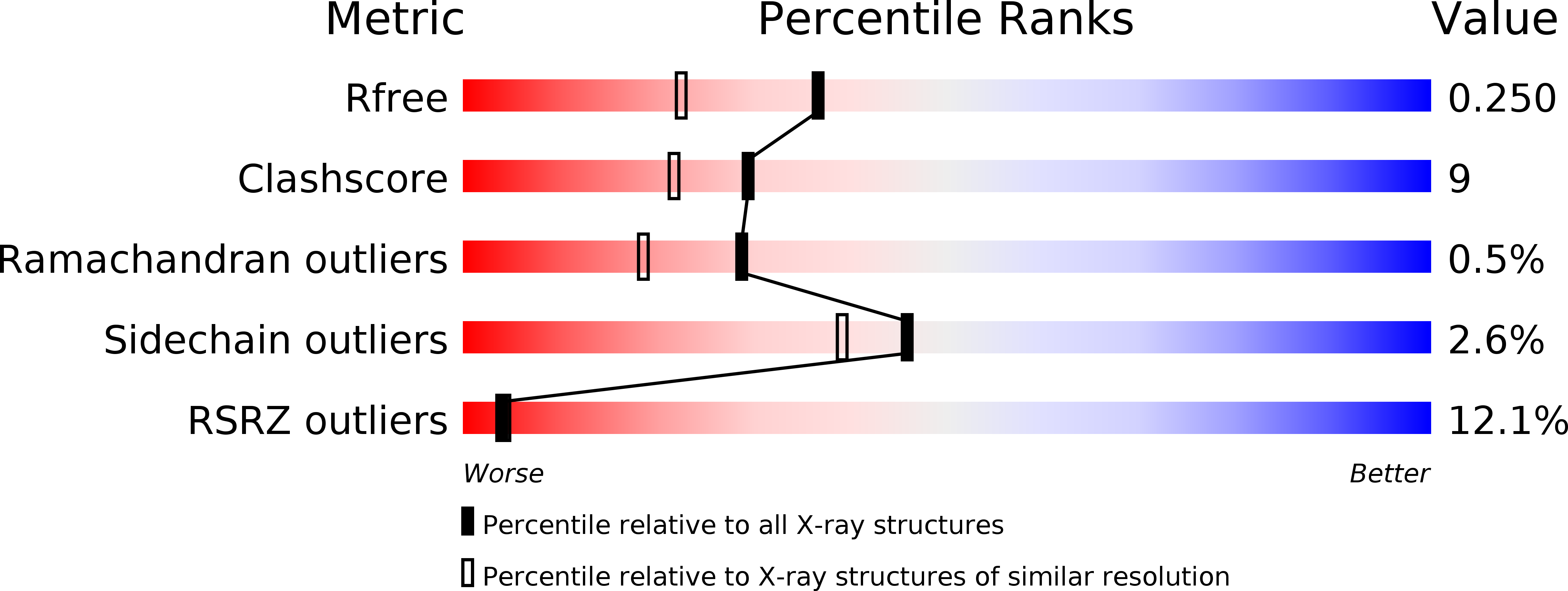
2PF5 image
Deposition Date 2007-04-04
Release Date 2007-06-26
Last Version Date 2024-10-30
Entry Detail
PDB ID:
2PF5
Keywords:
Title:
Crystal Structure of the Human TSG-6 Link Module
Biological Source:
Source Organism:
Homo sapiens (Taxon ID: 9606)
Method Details:
Experimental Method:
Resolution:
1.90 Å
R-Value Free:
0.22
R-Value Work:
0.19
R-Value Observed:
0.19
Space Group:
C 1 2 1
Macromolecular Entities
Structures with similar UniProt ID
Protein Blast
Polymer Type:polypeptide(L)
Molecule:Tumor necrosis factor-inducible protein TSG-6
Gene (Uniprot):TNFAIP6
Chain IDs:A, B, C, D, E
Chain Length:98
Number of Molecules:5
Biological Source:Homo sapiens
Primary Citation
Plasticity of the TSG-6 HA-binding loop and mobility in the TSG-6-HA complex revealed by NMR and X-ray crystallography
J.Mol.Biol. 371 669 684 (2007)
PMID: 17585936 DOI: 10.1016/j.jmb.2007.05.073

Abstact

Tumour necrosis factor-stimulated gene-6 (TSG-6) is a glycosaminoglycan-binding protein expressed during inflammatory and inflammation-like processes. Previously NMR structures were calculated for the Link module of TSG-6 (Link_TSG6) in its free state and when bound to an octasaccharide of hyaluronan (HA(8)). Heparin was found to compete for HA binding even though it interacts at a site that is distinct from the HA-binding surface. Here we present crystallography data on the free protein, and (15)N NMR relaxation data for the uncomplexed and HA(8)-bound forms of Link_TSG6. Although the Link module is comparatively rigid overall, the free protein shows a high degree of mobility in the beta4/beta5 loop and at the Cys47-Cys68 disulfide bond, both of which are regions involved in HA binding. When bound to HA(8), this dynamic behaviour is dampened, but not eliminated, suggesting a degree of dynamic matching between the protein and sugar that may decrease the entropic penalty of complex formation. A further highly dynamic residue is Lys54, which is distant from the HA-binding site, but was previously shown to be involved in heparin binding. When HA is bound, Lys54 becomes less mobile, providing evidence for an allosteric effect linking the HA and heparin-binding sites. A mechanism is suggested involving the beta2-strand and alpha2-helix. The crystal structure of free Link_TSG6 contains five molecules in the asymmetric unit that are highly similar to the NMR structure and support the dynamic behaviour seen near the HA-binding site: they show little or no electron density for the beta4/beta5 loop and display multiple conformations for the Cys47-Cys68 disulfide bond. The crystal structures were used in docking calculations with heparin. An extended interface between a Link_TSG6 dimer and heparin 11-mer was identified that is in excellent agreement with previous mutagenesis and calorimetric data, providing the basis for further investigation of this interaction.

Legend

Protein

Chemical

Disease

Primary Citation of related structures