2PES image
Deposition Date 2007-04-03
Release Date 2008-04-22
Last Version Date 2024-02-21
Entry Detail
PDB ID:
2PES
Keywords:
Title:
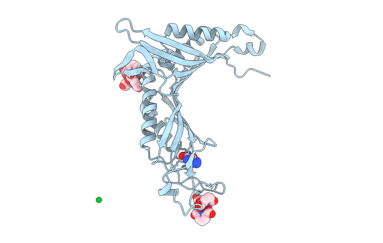
Urate Oxidase in complex with tris-dipicolinate Lutetium
Biological Source:
Source Organism:
Method Details:
Experimental Method:
Resolution:
1.60 Å
R-Value Free:
0.19
R-Value Work:
0.17
R-Value Observed:
0.18
Space Group:
I 2 2 2
Macromolecular Entities
Polymer Type:polypeptide(L)
Molecule:Uricase
Gene (Uniprot):uaZ
Chain IDs:A
Chain Length:295
Number of Molecules:1
Biological Source:Aspergillus flavus
Primary Citation
A dipicolinate lanthanide complex for solving protein structures using anomalous diffraction.
Acta Crystallogr.,Sect.D 66 762 769 (2010)
PMID: 20606256 DOI: 10.1107/S0907444910010954

Abstact

Tris-dipicolinate lanthanide complexes were used to prepare derivative crystals of six proteins: hen egg-white lysozyme, turkey egg-white lysozyme, thaumatin from Thaumatococcus daniellii, urate oxidase from Aspergillus flavus, porcine pancreatic elastase and xylanase from Trichoderma reesei. Diffraction data were collected using either synchrotron radiation or X-rays from a laboratory source. In all cases, the complex turned out to be bound to the protein and the phases determined using the anomalous scattering of the lanthanide led to high-quality electron-density maps. The binding mode of the complex was characterized from the refined structures. The lanthanide tris-dipicolinate was found to bind through interactions between carboxylate groups of the dipicolinate ligands and hydrogen-bond donor groups of the protein. In each binding site, one enantiomeric form of the complex is selected from the racemic solution according to the specific site topology. For hen egg-white lysozyme and xylanase, derivative crystals obtained by cocrystallization belonged to a new monoclinic C2 crystal form that diffracted to high resolution.

Legend

Protein

Chemical

Disease

Primary Citation of related structures