2PCX image
Deposition Date 2007-03-30
Release Date 2008-04-08
Last Version Date 2023-08-30
Entry Detail
PDB ID:
2PCX
Keywords:
Title:
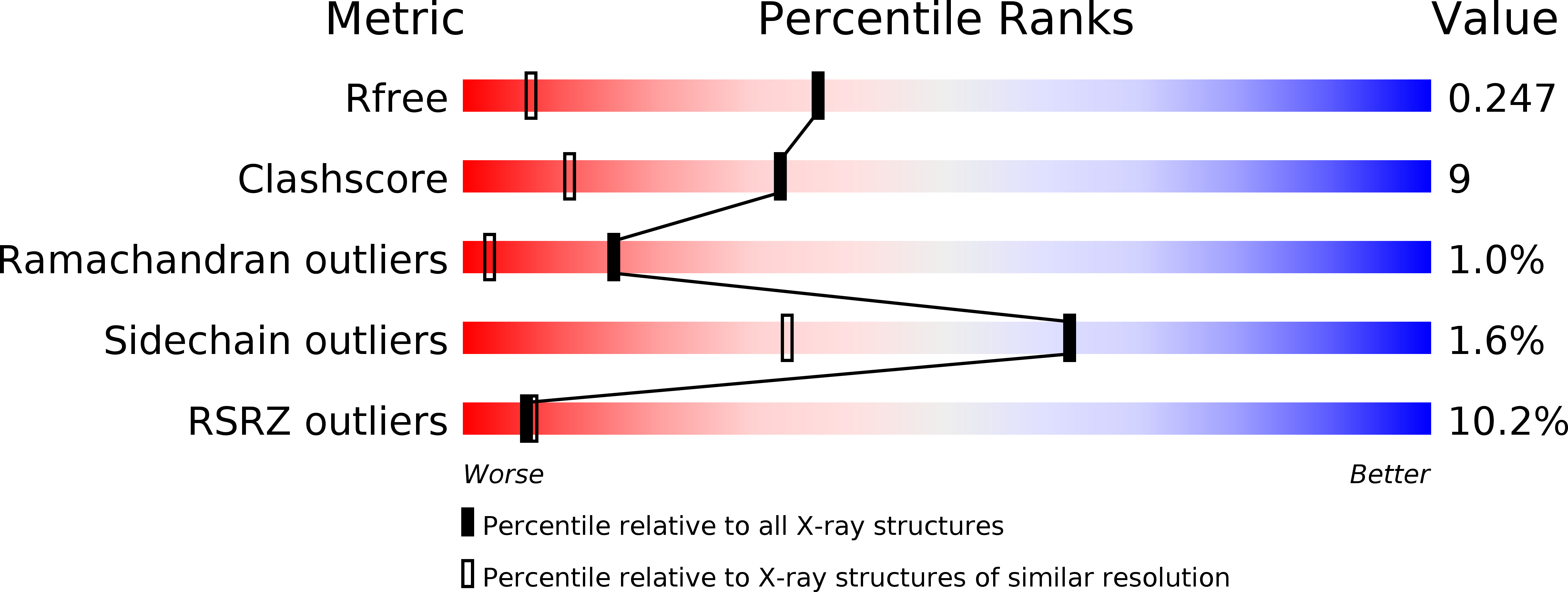
Crystal structure of p53DBD(R282Q) at 1.54-angstrom Resolution
Biological Source:
Source Organism:
Homo sapiens (Taxon ID: 9606)
Method Details:
Experimental Method:
Resolution:
1.54 Å
R-Value Free:
0.23
R-Value Work:
0.19
R-Value Observed:
0.19
Space Group:
P 1
Macromolecular Entities
Polymer Type:polypeptide(L)
Molecule:Cellular tumor antigen p53
Gene (Uniprot):TP53
Mutations:R282Q
Chain IDs:A
Chain Length:220
Number of Molecules:1
Biological Source:Homo sapiens
Ligand Molecules
Primary Citation
Impact of low-frequency hotspot mutation R282Q on the structure of p53 DNA-binding domain as revealed by crystallography at 1.54 angstroms resolution.
Acta Crystallogr.,Sect.D 64 471 477 (2008)
PMID: 18453682 DOI: 10.1107/S0907444908003338

Abstact

Tumor suppressor p53 is a sequence-specific DNA-binding protein and its central DNA-binding domain (DBD) harbors six hotspots (Arg175, Gly245, Arg248, Arg249, Arg273 and Arg282) for human cancers. Here, the crystal structure of a low-frequency hotspot mutant, p53DBD(R282Q), is reported at 1.54 angstroms resolution together with the results of molecular-dynamics simulations on the basis of the structure. In addition to eliminating a salt bridge, the R282Q mutation has a significant impact on the properties of two DNA-binding loops (L1 and L3). The L1 loop is flexible in the wild type, but it is not flexible in the mutant. The L3 loop of the wild type is not flexible, whereas it assumes two conformations in the mutant. Molecular-dynamics simulations indicated that both conformations of the L3 loop are accessible under biological conditions. It is predicted that the elimination of the salt bridge and the inversion of the flexibility of L1 and L3 are directly or indirectly responsible for deactivating the tumor suppressor p53.

Legend

Protein

Chemical

Disease

Primary Citation of related structures