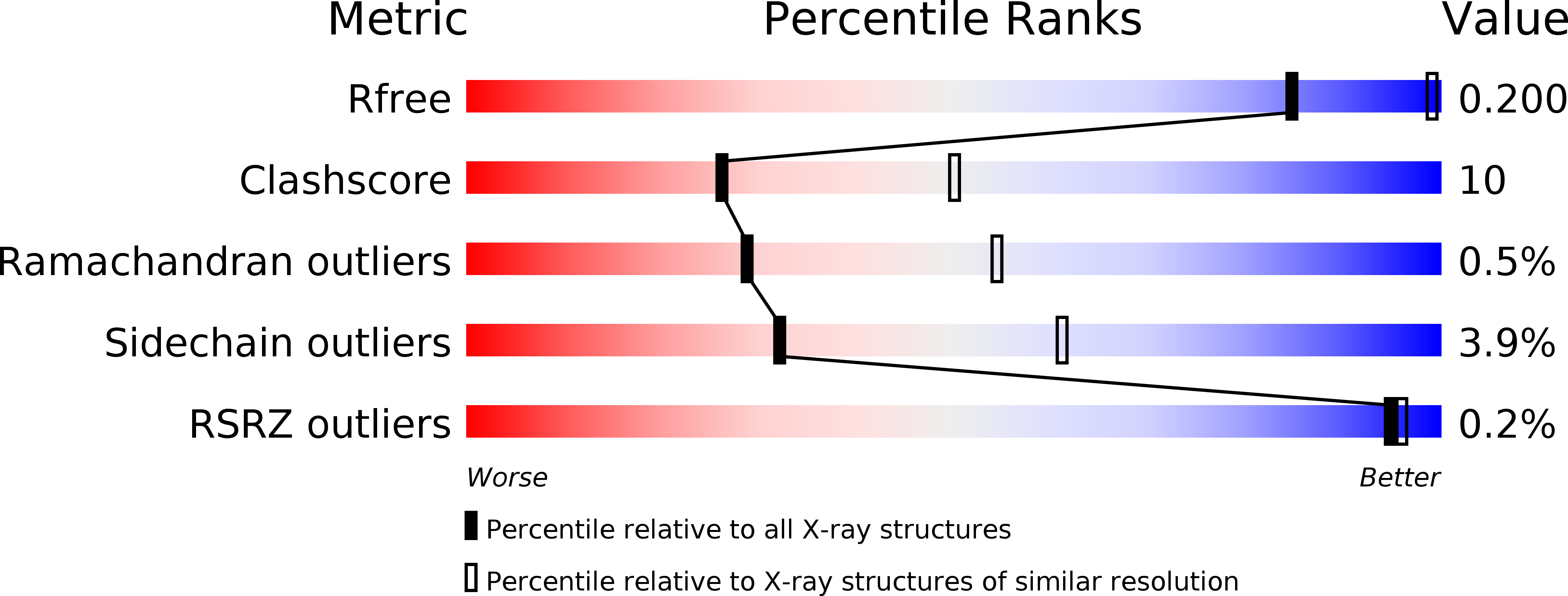
2PAA image
Deposition Date 2007-03-27
Release Date 2007-11-27
Last Version Date 2023-08-30
Entry Detail
PDB ID:
2PAA
Keywords:
Title:
Crystal structure of phosphoglycerate kinase-2 bound to atp and 3pg
Biological Source:
Source Organism:
Mus musculus (Taxon ID: 10090)
Host Organism:
Method Details:
Experimental Method:
Resolution:
2.70 Å
R-Value Free:
0.27
R-Value Work:
0.19
Space Group:
P 1
Macromolecular Entities
Polymer Type:polypeptide(L)
Molecule:Phosphoglycerate kinase, testis specific
Gene (Uniprot):Pgk2
Chain IDs:A, B
Chain Length:416
Number of Molecules:2
Biological Source:Mus musculus
Primary Citation
X-ray analysis of phosphoglycerate kinase 2, a sperm-specific isoform from Mus musculus.
Proteins 71 1134 1144 (2007)
PMID: 18004764 DOI: 10.1002/prot.21801

Abstact

Phosphoglycerate kinase 2 (PGK2) is an isozyme of the glycolytic pathway that provides ATP required for sperm motility. It is encoded by an autosomal retrogene that is expressed only during spermatogenesis, concomitant with the inactivation of the X-linked Pgk1 gene. PGK2 from the mouse, Mus musculus, has been overexpressed from a plasmid in bacteria and purified. It was crystallized in three forms: as the apoenzyme, as a complex with 3-phosphoglycerate (3PG), and as a complex with 3PG and ATP. The crystal structures were solved to 2.7, 2.0, and 2.7 A resolutions, respectively. The overall fold is nearly identical with previously solved mammalian PGK1 molecules. The apoenzyme is in the "open" form; that is the N-terminal domain that can bind 3PG and the C-terminal domain that binds ATP are too far apart for the substrates to interact. Binding 3PG causes a 13 degree rotation that partially closes the structure and causes helix 13, which is disordered in the unliganded structure, to stabilize. Binding ATP leaves the protein in the open configuration but also causes helix 13 to be ordered. Sequence alignment suggests that the active site of PGK2 is essentially identical to that of the cytoplasmic PGK1, but significant differences accumulate on a side of the C-terminal domain away from the active site. These changes may mediate the binding of this isoform to other proteins within the sperm flagellum, while still allowing the hinging action between the domains that is essential to catalytic activity.

Legend

Protein

Chemical

Disease

Primary Citation of related structures