
Deposition Date
2007-03-20
Release Date
2007-07-17
Last Version Date
2023-08-30
Entry Detail
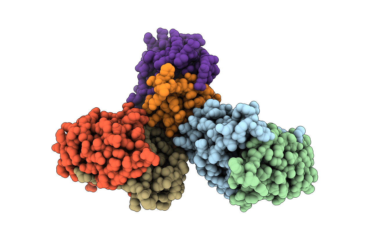
PDB ID:
2P7M
Keywords:
Title:
Crystal structure of monoclinic form of genomically encoded fosfomycin resistance protein, FosX, from Listeria monocytogenes at pH 6.5
Biological Source:
Source Organism(s):
Listeria monocytogenes (Taxon ID: 169963)
Expression System(s):
Method Details:
Experimental Method:
Resolution:
1.85 Å
R-Value Free:
0.26
R-Value Work:
0.23
Space Group:
C 1 2 1
