
Deposition Date
2007-03-19
Release Date
2007-08-21
Last Version Date
2024-02-21
Entry Detail
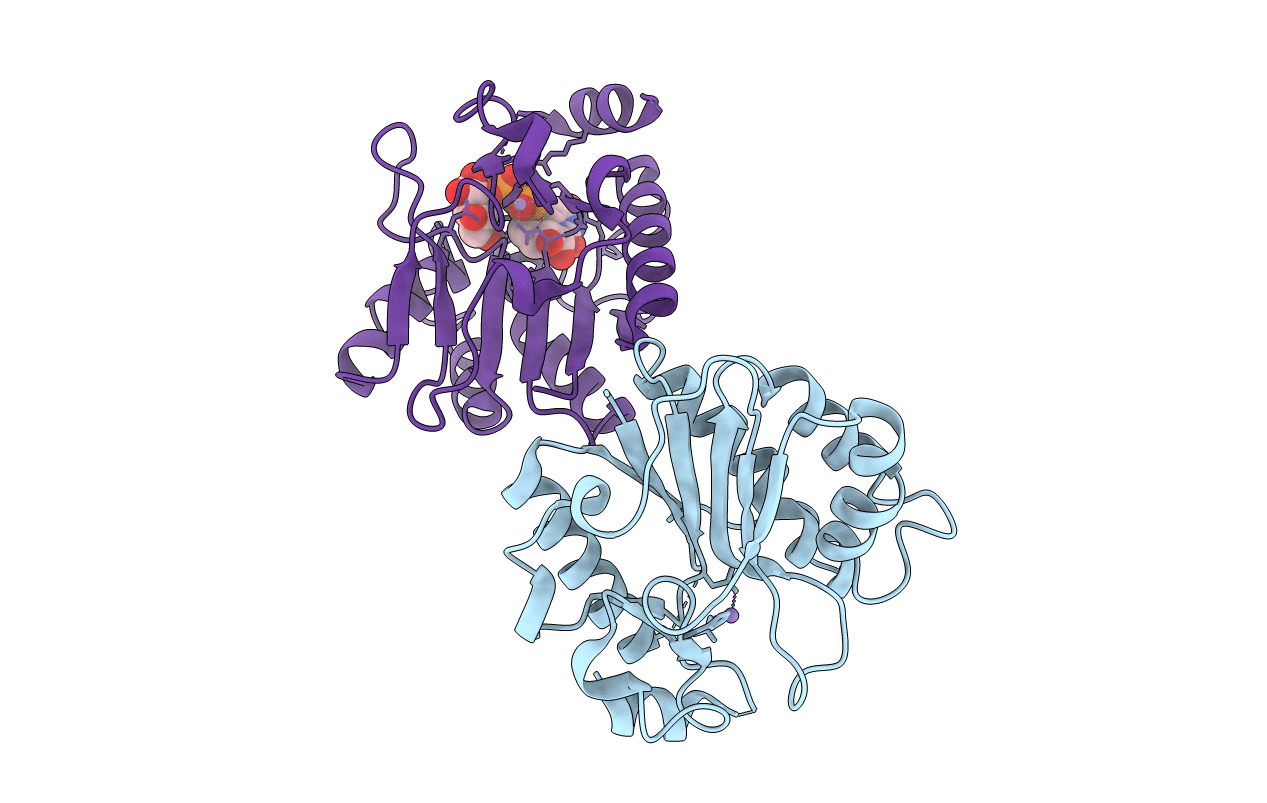
PDB ID:
2P72
Keywords:
Title:
crystal structure of a glycosyltransferase involved in the glycosylation of the major capsid of PBCV-1
Biological Source:
Source Organism(s):
Paramecium bursaria Chlorella virus 1 (Taxon ID: 10506)
Expression System(s):
Method Details:
Experimental Method:
Resolution:
2.00 Å
R-Value Free:
0.23
R-Value Work:
0.20
R-Value Observed:
0.20
Space Group:
C 1 2 1
