Deposition Date
2007-03-16
Release Date
2007-04-03
Last Version Date
2024-11-06
Entry Detail
PDB ID:
2P6A
Keywords:
Title:
The structure of the Activin:Follistatin 315 complex
Biological Source:
Source Organism(s):
Homo sapiens (Taxon ID: 9606)
Expression System(s):
Method Details:
Experimental Method:
Resolution:
3.40 Å
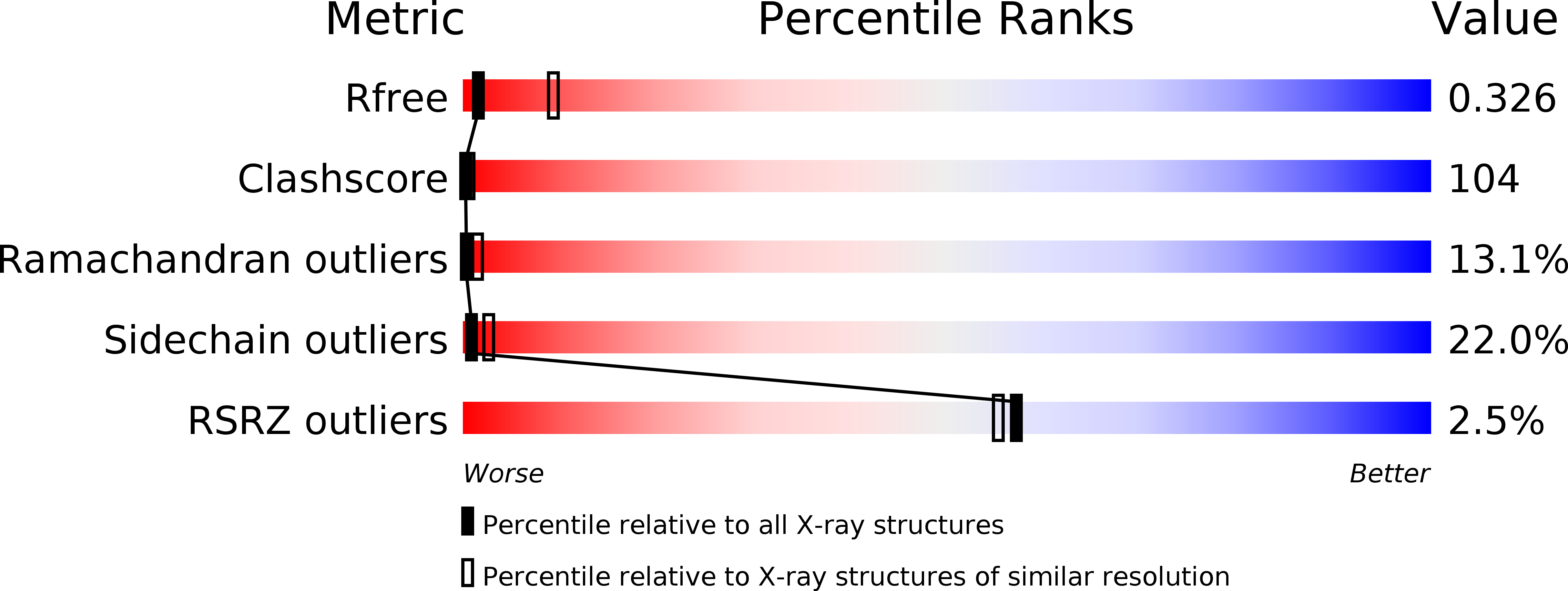
R-Value Free:
0.32
R-Value Work:
0.22
R-Value Observed:
0.22
Space Group:
P 2 2 21