2P66 image
Deposition Date 2007-03-16
Release Date 2007-04-10
Last Version Date 2023-08-30
Entry Detail
PDB ID:
2P66
Title:
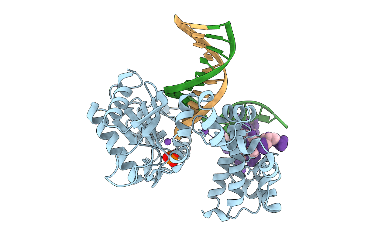
Human DNA Polymerase beta complexed with tetrahydrofuran (abasic site) containing DNA
Biological Source:
Source Organism:
Homo sapiens (Taxon ID: 9606)
Host Organism:
Method Details:
Experimental Method:
Resolution:
2.50 Å
R-Value Free:
0.28
R-Value Work:
0.21
R-Value Observed:
0.21
Space Group:
P 1 21 1
Macromolecular Entities
Structures with similar UniProt ID
Protein Blast
Polymer Type:polypeptide(L)
Molecule:DNA polymerase beta
Gene (Uniprot):POLB
Chain IDs:D (auth: A)
Chain Length:335
Number of Molecules:1
Biological Source:Homo sapiens
Primary Citation
Structural insight into the DNA Polymerase beta deoxyribose phosphate lyase mechanism
DNA REPAIR 4 1347 1357 (2005)
PMID: 16172026 DOI: 10.1016/j.dnarep.2005.08.009

Abstact

A large number of biochemical and genetic studies have demonstrated the involvement of DNA polymerase beta (Pol beta) in mammalian base excision repair (BER). Pol beta participates in BER sub-pathways by contributing gap filling DNA synthesis and lyase removal of the 5'-deoxyribose phosphate (dRP) group from the cleaved abasic site. To better understand the mechanism of the dRP lyase reaction at an atomic level, we determined a crystal structure of Pol beta complexed with 5'-phosphorylated abasic sugar analogs in nicked DNA. This DNA ligand represents a potential BER intermediate. The crystal structure reveals that the dRP group is bound in a non-catalytic binding site. The catalytic nucleophile in the dRP lyase reaction, Lys72, and all other potential secondary nucleophiles, are too far away to participate in nucleophilic attack on the C1' of the sugar. An approximate model of the dRP group in the expected catalytic binding site suggests that a rotation of 120 degrees about the dRP 3'-phosphate is required to position the epsilon-amino Lys72 close to the dRP C1'. This model also suggests that several other side chains are in position to facilitate the beta-elimination reaction. From results of mutational analysis of key residues in the dRP lyase active site, it appears that the substrate dRP can be stabilized in the observed non-catalytic binding conformation, hindering dRP lyase activity.

Legend

Protein

Chemical

Disease

Primary Citation of related structures
Feedback Form
Name
Email
Institute
Feedback