Deposition Date
2007-03-16
Release Date
2007-03-27
Last Version Date
2023-08-30
Entry Detail
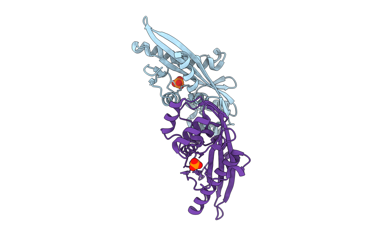
PDB ID:
2P5X
Keywords:
Title:
Crystal structure of Maf domain of human N-acetylserotonin O-methyltransferase-like protein
Biological Source:
Source Organism(s):
Homo sapiens (Taxon ID: 9606)
Expression System(s):
Method Details:
Experimental Method:
Resolution:
2.00 Å
R-Value Free:
0.25
R-Value Work:
0.19
R-Value Observed:
0.20
Space Group:
P 21 21 2